Keloid Guidelines: BMJ Best Practice (2024)
Keloid is a form of abnormal scarring that occurs in susceptible individuals. Diagnosis is clinical. Treatment options include injectable preparations, cryotherapy, as well as surgery and radiation therapy.
In this article
- Summary
- Definition
- Epidemiology
- Etiology
- Pathophysiology
- Case history
- Case history #1
- Other presentations
- Approach
- History and exam
- Risk factors
- Tests
- Differentials
- Approach
- Treatment algorithm overview
- Treatment algorithm
- Emerging
- Primary prevention
- Patient discussions
- Monitoring
- Complications
- Prognosis
- Diagnostic guidelines
- Treatment guidelines
- Key articles
- References
BMJ Best Practice topic: Keloid. Last updated: Oct 08, 2024. © BMJ Publishing Group Ltd 2024. All rights reserved.
Summary
Keloid is a form of abnormal scarring that occurs in susceptible individuals.
More common in darker skin types.
Diagnosis is clinical.
Treatment options include injectable preparations, cryotherapy, as well as surgery and radiation therapy.
Definition
Keloid scarring can be defined using a variety of clinical and histologic parameters. The most widely accepted definition relates to bulky scar formation, which extends beyond the margins of the original injury to the skin and shows lack of spontaneous regression. Histologically, a keloid represents a fibroproliferative disorder within the reticular dermis, characterized by sustained inflammation, angiogenesis, and collagen accumulation organized in hyalinized form.
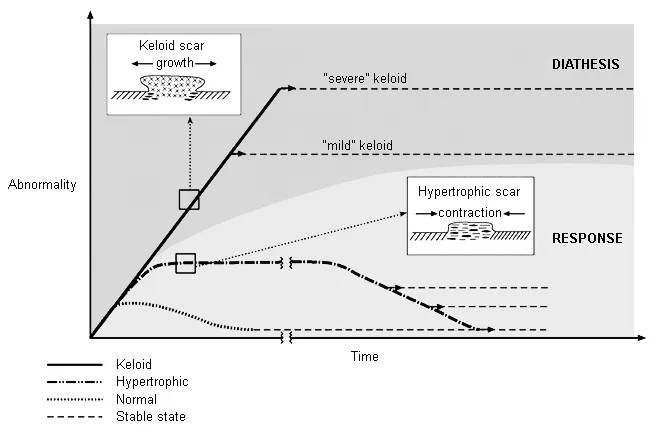
These clinical and histologic features differ from hypertrophic scars, which represent bulky scars limited within the confines of the original injury to the skin; for a large number of individuals, hypertrophy represents a transient stage of scar maturation, which settles with time. Histologically, hypertrophic scars are characterized by dermal nodule formation as opposed to hyalinization. Keloid scars are increasingly considered an extreme form of hypertrophic scarring based on both clinical and histologic evolution; in other words, a hypertrophic stage precedes the development of a keloid scar.[1]
This topic focuses on keloids in adults. Management of keloids in children is more challenging and beyond the scope of this topic.
Epidemiology
Keloids are only found in humans and occur in 1% to 16% of the population, with significant ethnicity-related differences.[2] Keloids are most common among Afro-Caribbean people, with an estimated incidence of 5% to 10%.[3] Some studies report an increased prevalence in Asian people compared with white people.[4] [5] However, the reported incidence is similar in Japan (0.1%) and England (0.09%).[3]
It is thought that men and women are equally affected. The average age at onset is 10-30 years.[6]
Etiology
Keloid scarring usually develops in response to inciting inflammation or trauma to the dermis. Common inciting events include:[7] [8]
- Acne
- Body piercing
- Tattoos
- Insect bites
- Vaccinations (e.g., BCG)
- Surgery
- Follicular inflammation
Keloids typically occur in patients with darker skin types, suggesting a potential role of melanin and/or melanocytes in scarring. However, one survey in Kenya and Tanzania reported a similar prevalence of keloids in people with albinism.[9]
Numerous patients with keloid scars report a positive family history of pathologic scarring, which may suggest that there are genetic factors implicated in the pathophysiology. Studies of familial keloid cases have identified associated single nucleotide polymorphisms and susceptibility loci on chromosomes 1q41, 2q23, 7p11, and 10q23.31 in Japanese, African-American, and Han Chinese families.[10] [11] [12] Responsible genes have not yet been identified.
Growth factors are also thought to have a role. There is an observed resistance to apoptosis in keloid fibroblasts, which may contribute to the slowly progressive and long-lasting nature of the lesions.[13] Growth factors associated with the pathogenesis of keloid scarring include connective tissue growth factor (CTGF) and platelet-derived growth factor (PDGF). A number of other growth factors are under investigation.[14] [15]
Hormones may also play a part, given the observation that keloid scars often develop or worsen at puberty and during pregnancy, and that there is often an improvement after menopause.[16] Estrogen has vasodilatory action, and hence may enhance levels of inflammation.[17]
Pathophysiology
The exact pathophysiology of keloids remains unclear. In part, this is due to the heterogeneous nature of the scar and the multifactorial nature of the possible etiologic associations.[18]
The scar arises from excessive dermal collagen deposition, with more cellular activity at the periphery than in the center; central cells demonstrate a decreased tendency to undergo apoptosis. The extracellular matrix and vascular architecture of keloid scars varies within each scar and between scars, and is distinctly different in the spectrum of heterogeneity from that of hypertrophic scarring.
Keloid is a unique form of scarring because it does not pass through a cycle of proliferation, stabilization, and involution. Rather, it proliferates and then stabilizes. However, the point of stabilization and consequent size varies considerably, both between and within patients. Unlike hypertrophic scarring, keloids are not typically associated with contractures.
Case History
Case History #1
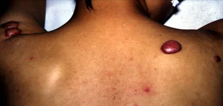
An 18-year-old woman with a history of acne presents with a 20 × 15 mm raised, firm, pink swelling on her right upper back. This has been slowly growing for more than 1 year. She has also noticed a new smaller swelling, also pink and firm, developing in the adjacent skin. She already has 2 similar lesions, which developed on her left upper arm after vaccinations she received when she entered high school. These grew for about 2 years, but there has been no subsequent change in their size. Occasionally, they itch or she experiences a sharp, needle-like pain. She has never sought treatment before but is now self-conscious as she is hoping to wear a low-cut dress for her older sister's wedding.
Other Presentations
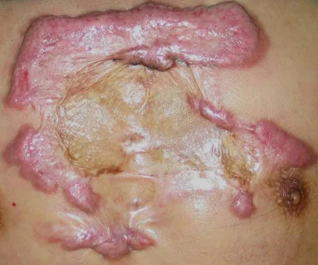
Typically, keloid scarring occurs after an inciting event such as trauma, surgery, body piercing, vaccination (in the deltoid region), or acne on the back or chest. However, it may occur on any part of the body in susceptible individuals.
Approach
The diagnosis of keloid scarring is primarily clinical. A skin biopsy is rarely required and must be weighed against the risk of making the condition worse.
Clinical History
Unlike hypertrophic scarring, keloid is not typically associated with skin contractures.
Typical findings include:[19]
- An inciting event (e.g., body piercing, surgery, or trauma in a predisposed individual)
- Personal or family history of keloids
- High-risk skin type
- Symptoms of itch and pain
- Gradual increase in size of lesions
- Lack of spontaneous regression.
Physical Exam
Site
- Typically located over high tension bodily areas (e.g., upper back/deltoid, presternal, lower abdominal regions) and over bony prominences.
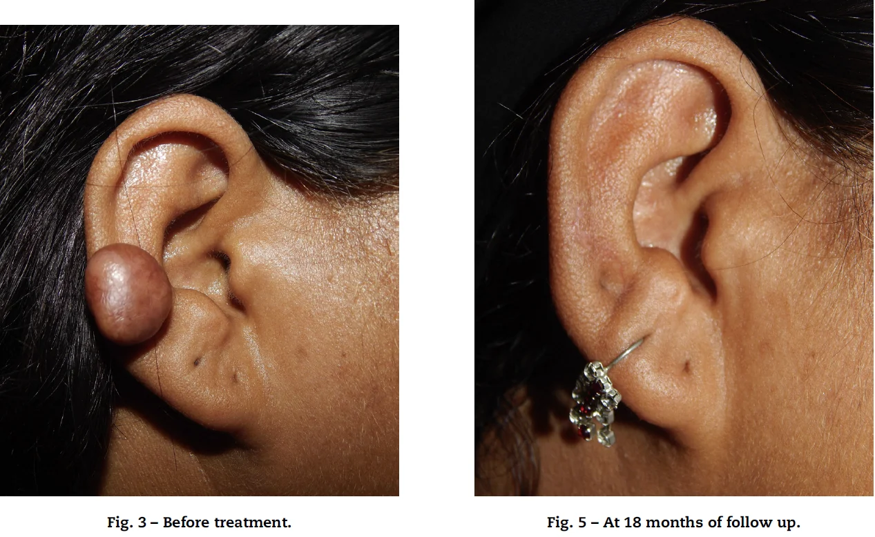
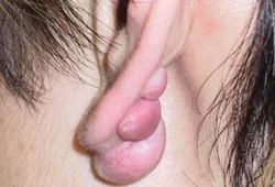
- Earlobes are also a common location, especially after ear piercing.[19]
Texture or Surface
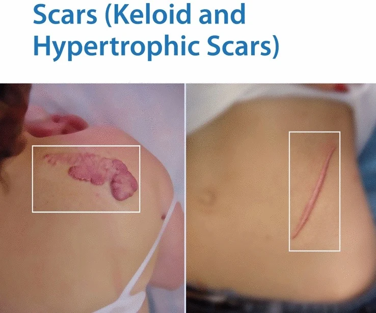
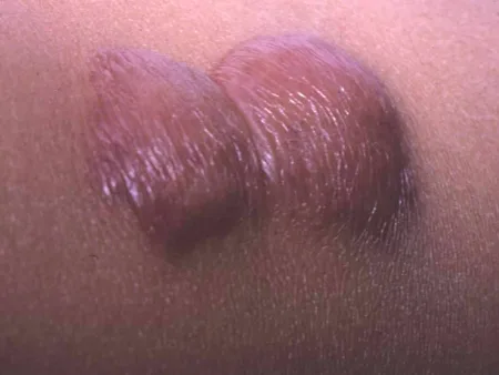
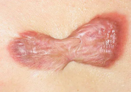
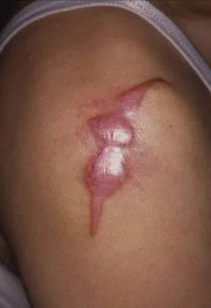
- The surface of the scar is typically smooth and shiny with an overhanging edge.
Consistency
- Most keloid scars tend to be firm in consistency, especially if they are long-standing.
Color
- Keloid scars may exhibit a range of colors depending on the normal skin pigmentation. During the active-growth phase, the scar may be erythematous, indicating increased blood flow. As the growth slows and begins to stabilize, the keloid may become more similar in color to the surrounding skin.
Investigations
Diagnosis is based on clinical findings.[17] [19] The main reason to perform a skin biopsy is when there is doubt about the nature of the lesion or clinical suspicion of malignancy (e.g., rapid growth).[21]
The most important differential diagnosis is dermatofibrosarcoma, which typically involves more subdermal spread (the term subdermal is used rather than subcutaneous to underscore the pattern of growth). Clinically, the skin can appear to be normal on inspection but has a palpable tumor that arises from the dermis and spreads into the underlying tissue.
The need for histologic exam following keloidectomy should be discussed with individual patients. The risk of misdiagnosis of other pathology as keloid scar has been estimated to be 1.06%.[22]
History and Exam
Key Diagnostic Factors
History of Inciting Event (e.g., Body Piercing, Surgery, Vaccination) (Common)
- Typically there is an inciting event that has caused damage to the skin.[1] There may be an interval of months between the damage and the first appearance of the scar.
- Occasionally, there will be no clear history of an inciting event.
Slow Growth/lack of Spontaneous Regression (Common)
- Keloids are typically slow-growing lesions and fail to regress in most cases.
Erythematous, Smooth, and Shiny Elevated Scar With Overhanging Edge (Common)
- Typically, the keloid scar grows beyond the boundaries of the original injury.
Other Diagnostic Factors
Itch or Pain (Uncommon)
- These symptoms tend to be more intense during the active growth phase.
Risk Factors
Strong
History of Prior Keloid
- A history of prior keloid scarring in a patient is associated with a strong risk of occurrence at other sites.[19]
- Research on the differential gravity of risk factors for keloid development is limited and inconsistent, and thus the classification into strong and weak may change as new evidence emerges.[3]
Local Skin Tension
- Keloids occur most frequently in areas of the body subject to stretch including the upper back/shoulders, chest, and lower abdomen.[20]
- Research on the differential gravity of risk factors for keloid development is limited and inconsistent, and thus the classification into strong and weak may change as new evidence emerges.[3]
Follicular Inflammation/infection
- If the skin gets rewounded, becomes infected, or consistently inflamed during healing, the risk of keloid scar formation is higher by virtue of a prolonged inflammatory response.[20]
- Research on the differential gravity of risk factors for keloid development is limited and inconsistent, and thus the classification into strong and weak may change as new evidence emerges.[3]
Family History of Keloid and Genetic Factors
- Numerous patients with keloid scars report a positive family history of pathologic scarring, which may suggest that there are genetic factors implicated in the pathophysiology. Studies of familial keloid cases have identified associated single nucleotide polymorphisms and susceptibility loci on chromosomes 1q41, 2q23, 7p11, and 10q23.31 in Japanese, African-American, and Han Chinese families.[10] [11] [12] Responsible genes have not yet been identified.
- Research on the differential gravity of risk factors for keloid development is limited and inconsistent, and thus the classification into strong and weak may change as new evidence emerges.[3]
Weak
Dark Skin Types
- Keloids are most common among Afro-Caribbean people.[3] Some studies report an increased prevalence in Asian people compared with white people.[4] [5]
- Research on the differential gravity of risk factors for keloid development is limited and inconsistent, and thus the classification into strong and weak may change as new evidence emerges.[3]
Hormonal Status
- Keloid scars often develop or worsen at puberty and during pregnancy, and an improvement is often noted after the menopause.[16] Estrogen has vasodilatory action, and hence may enhance levels of inflammation.[17]
- Research on the differential gravity of risk factors for keloid development is limited and inconsistent, and thus the classification into strong and weak may change as new evidence emerges.[3]
Systemic Conditions (e.g., Hypertension, Atopic Eczema, Castleman Disease)
- Keloids have been associated with a number of systemic conditions, including hypertension, atopic eczema, and conditions related to increased levels of cytokines such as Castleman disease.[4] [17] An analysis of UK Biobank data found a statistically significant association between excessive scarring and comorbid atopic eczema in Asian, white, and black patients.[4]
- Research on the differential gravity of risk factors for keloid development is limited and inconsistent, and thus the classification into strong and weak may change as new evidence emerges.[3]
Lifestyle Factors
- Several lifestyle parameters may exacerbate preexisting scars or surgery-induced inflammation, including vigorous physical activity (e.g., competitive athletes, manual workers).[17]
- Research on the differential gravity of risk factors for keloid development is limited and inconsistent, and thus the classification into strong and weak may change as new evidence emerges.[3]
Tests
1st Test to Order
Clinical Diagnosis
Result: features of keloid
- Usually no tests are necessary.
Other Tests to Consider
Skin Biopsy
Result: elevated cellular proliferation at the margins of the scar but reduced cell density within the central portions; may also demonstrate increased extracellular matrix deposition; collagen fibers are typically larger and more irregular in arrangement than in normal scars; presence of hyalinized collagen in keloids
- Rarely required and must be weighed against the risk of making the condition worse. The main reason to perform a skin biopsy is when there is doubt about the nature of the lesion (e.g., malignancy).[21] The most important differential diagnosis is dermatofibrosarcoma.
- The need for histologic exam following keloidectomy should be discussed with individual patients. The risk of misdiagnosis of other pathology as keloid scar has been estimated to be 1.06%.[22]
Differentials
Hypertrophic Scar
Differentiating Signs / Symptoms
- No association with skin type.
- Always preceded by injury.
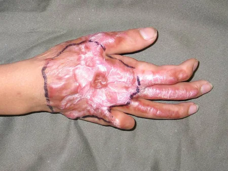
- On physical exam there may be evidence contractures.
Differentiating Tests
- Diagnosis is clinical.
- Immunohistochemistry demonstrates flatter, less distinct bundles of fibers, presence of myofibroblasts, and increased density of blood vessels and cells; dermal nodules are present.
Dermatofibrosarcoma Protuberans
Differentiating Signs / Symptoms
- Clinically, often starts as an asymptomatic papule or nodule, but the skin can appear to be normal on inspection. The skin lesion is contiguous with a palpable tumor that arises from the dermis and spreads into the underlying tissue.
Differentiating Tests
- Diagnosis may be difficult; histologic exam is necessary when there is doubt. This demonstrates neoplastic spindle-shaped nuclei.
- An incisional biopsy with a large sample is required.
Dermatofibroma
Differentiating Signs / Symptoms
- Usually, no precipitating trauma but similar slow growth.
- Clinically, may appear as raised skin nodule but may have subdermal extension.
Differentiating Tests
- Skin biopsy demonstrates proliferation of fibroblasts and dermal fibrosis.
Giant Cell Fibroblastoma
Differentiating Signs / Symptoms
- Clinically presents as a firm irregular nodule, which may be tender on palpation. Affects mostly prepubertal children with the trunk being the most common location.[23] Although locally aggressive, metastases have not been reported.
Differentiating Tests
- Biopsy demonstrates a nodular mass composed of spindle cells with scattered giant cells occupying most of the reticular dermis.
Approach
The existing evidence to support the ideal treatment of keloids is weak.[24] [25] A number of review articles have been published summarizing consensual algorithms for keloid scar management, with most of them emphasizing the need for combined multidisciplinary therapeutic regimens.[17] [26] This is pertinent given the wide heterogeneity of scar features, as well as the different therapeutic goals and objectives that patients present with.[17] [27]
Initial Treatment
The first-line treatment will depend on the site, size, etiology, and history of previous treatments. In general terms, single, small keloid scars can be treated with nonsurgical modalities, whereas large, bulky, or multiple lesions should be assessed for surgery and adjuvant therapy.[17] [28] Patient preference is a strong determining factor as well, so the number and size of presenting scarring should not preclude consideration of surgery.[17]
Nonsurgical local therapy may include corticosteroids, antitumor agents, laser therapy, or cryotherapy. Treatments may be combined or used sequentially to improve efficacy and minimize adverse effects. Examples of combinations include intralesional fluorouracil plus corticosteroid injections, laser therapy plus corticosteroid injections, or laser therapy plus intralesional fluorouracil. Preferred options are not well defined due to limited evidence. Take an individualized approach after discussing treatment options and goals with the patient.
See also Patient discussions .
Corticosteroids
Corticosteroids can be administered as an intralesional or sublesional injection or as a topically applied tape, and may be used in combination with other treatments. Corticosteroid tapes can be used as monotherapy in patients with thin scars. Bulkier lesions require a more reinforced approach, involving either induction with injectables or combination with other modalities. Benefits of a combined approach include enhanced efficacy and minimization of corticosteroid-associated adverse effects.[29] [30]
Corticosteroids have a number of different actions including:[31] [32]
- Inhibition of leukocyte/monocyte migration and phagocytosis.
- Vasoconstriction which reduces the delivery of oxygen and nutrients to the skin/scar.
- Antimitotic effect through inhibition of fibroblast proliferation (by virtue of decreasing mitogenic factors including transforming growth factor beta-1 [TGF-beta 1], vascular endothelial growth factor, and insulin-like growth factor-1) and induction of cellular degeneration.
- Decrease of collagen synthesis and enhancement of collagen degradation; this occurs through the reduction of levels of alpha-2 macroglobulin and alpha-1 antitrypsin (which are both potent collagenase inhibitors).
Corticosteroid Injections
The most commonly used injectable corticosteroid is triamcinolone (as the acetonide salt). Despite the popularity of corticosteroid injections for the treatment of keloid and hypertrophic scars, little conclusive evidence exists to support definitive treatment protocols. The frequency of administration varies widely in the literature and ranges from 1- to 8-week intervals between each injectable treatment.[33] [34] A survey among 102 international plastic surgeons concluded that the most popular interval between injections is 3-4 weeks (31%), closely followed by 4-5 weeks (26%).[35] Some proposed protocols also recommended using different doses depending on the volume/dimensions as well as location of the scar.[36] [37] [38] Response rates to triamcinolone injections vary considerably from 50% to 100% regression; recurrence rates are estimated to be 33% at year 1 posttreatment and 50% after 5 years.[33]
- Intralesional injection: the most popular technique for injection into the substance of the keloid. Small diameter needles (ranges between 25 and 30 Gauge) in conjunction with Luer lock or insulin syringes with an integrated needle have been advocated in order to withstand the pressure encountered during injection into stiff scars. Several authors have advised that the needle should be inserted into the papillary dermis (where collagenase is produced) and the substance administered while slowly withdrawing the needle, while others support targeting the mid-dermis level in order to avoid irreversible epidermal atrophy, which is more likely with superficial injection. The principle of inserting the needle within the keloidal lesion (as opposed to neighboring healthy skin) to avoid the development of an iatrogenic keloid has not been substantiated with evidence in the literature.
- Sublesional injection: an alternative technique to deliver corticosteroid to a keloid scar at the level of deep dermis in an attempt to mitigate the shortcomings of the intralesional technique. The first publication on this method employed light and electron microscope exam and showed that the positive changes in histologic appearance appeared earlier compared to those achieved with the intralesional approach with a similar pattern of adverse effects including atrophy, depigmentation, and telangiectasia.
- A number of different techniques can be employed to minimize discomfort, including local anesthetic creams or injections before corticosteroid administration. Some clinicians add lidocaine to the corticosteroid to give a 50/50 dilution. Lidocaine has a local anesthetic effect but will not reduce the pain of the injection (though it may make the subsequent pain more tolerable). It is also thought that lidocaine has local effects on fibroblasts, causing reduced collagen production and inhibiting cell proliferation.[39] [40]
- Complications of intralesional corticosteroid injections are common (reported in up to 63%) and include skin thinning, atrophy of the subdermal fat, telangiectasia, plaque formation, and pigmentary changes.[35] Systemic adverse effects are possible and can lead to adrenal suppression and Cushing syndrome. One systematic review confirmed 18 cases of Cushing syndrome after intralesional triamcinolone administration; 80% of cases occurred in children, and most after administration of more than 40 mg of triamcinolone within a period of 1 month.[35] Systemic hormone imbalance may also develop, which can result in water retention and menstrual irregularities in susceptible women.[24]
Corticosteroid Tape
Topically applied corticosteroid tape (e.g., flurandrenolide) is indicated for:[41]
- First-line treatment of small keloids
- Adjunctive treatment following either corticosteroid injection or surgical intervention
Once corticosteroid injections have reduced the thickness of the pathologic scar to a couple of millimeters, corticosteroid tape can be a patient-friendly way to continue corticosteroid delivery to the affected skin.
The delivery of a corticosteroid using a tape formulation was first described in the literature in 1967.[42] Percutaneous corticosteroid delivery acts through the combined effect of the barrier component, which creates a semiocclusive environment of controlled hydration, and the corticosteroid component, which has the well-known effect of hampering fibroblast proliferation and decreasing the amount of collagen in the scar. The two components act synergistically to enhance vasoconstriction within the scar.
The tape is applied to the index scar with minimal overlap to neighboring skin and needs to be changed regularly. Follow-up is recommended after an initial trial of 3 months in order to assess clinical response.[21]
One trial reported no systemic adverse effects after application of flurandrenolide, a moderate-potency corticosteroid tape, to scars up to 5% total body surface area for at least 6-8 weeks.[43] A separate observational study reported no systemic complications following at least 12 months of tape application in a mixed adult and pediatric cohort.[44]
Topical Corticosteroids
Evidence to support the use of topical corticosteroids (e.g., creams, ointments, lotions) as monotherapy for keloid scars is very weak, and they should not be recommended routinely.[45] In one prospective observational study, a triamcinolone lotion was applied on scars, with an adjacent part of the lesion left untreated to act as the comparison area. Assessment on a 4-point scale (including parameters for color, elevation, texture, itch, and pain) found a response rate of 44.4% for the group of 9 keloid patients after a maximum follow-up period of 7 months.[46] Another case series investigating the response to fluocinolone cream included 12 patients with keloids: 2 patients showed clearance, 7 showed significant response, and 3 showed poor or no response, with no author speculation about the reasons for the variable response.[47]
Antitumor Agents
Antitumor agents used for keloid treatment include fluorouracil, bleomycin, and mitomycin. Although some guidelines recommend antitumor agents as second-line options for refractory keloids, others suggest they should be considered as part of first-line therapy to help prevent recurrence.[28] [45]
Fluorouracil is a pyrimidine analog, which interferes with DNA synthesis by blocking thymidine. It is also thought to inhibit type I collagen gene expression and the effects of TGF-beta.[48] It is used as an intralesional injection in the management of keloids.
The clinical effectiveness of intralesional fluorouracil seems to be similar to corticosteroids, with recurrence rates of up to 47%.[49] One randomized controlled trial (RCT) compared intralesional fluorouracil to triamcinolone injections in 44 keloidal patients. Both treatments were equally effective in reducing keloid volume, but the adverse effects in the fluorouracil group (pain and superficial ulceration) were significantly worse.[50] In another comparative trial of 30 patient with keloids, triamcinolone injection significantly improved volume reduction compared with intralesional fluorouracil, and fluorouracil was associated with more adverse effects (although this difference was nonsignificant).[51]
The addition of intralesional fluorouracil to triamcinolone injection is believed to ameliorate the combined adverse effect profile because of the decreased dose requirement of each agent. In one meta-analysis of intralesional injections for hypertrophic and keloid scars, the combination of intralesional triamcinolone and fluorouracil injection was more effective than triamcinolone alone in terms of patient assessment, observer assessment, scar height after treatment, as well as erythema score.[30]
A very small number of studies have employed intralesional fluorouracil in combination with laser therapy or as a postoperative adjunct.[52] [53] [54] The consensus in the literature suggests that the addition of intralesional fluorouracil to other scar management modalities - including intralesional corticosteroids and pulse dye laser - improved efficacy versus each treatment alone.[29]
Bleomycin and mitomycin are alternative injectable treatments to flatten keloids, but not commonly used.
Bleomycin is used in the management of various malignancies and works by inducing tissue sclerosis. According to one meta-analysis, intralesional bleomycin significantly improves keloid and hypertrophic scars compared with triamcinolone, fluorouracil, and cryotherapy combined with triamcinolone.[55]
Mitomycin is a Streptomyces derivative that possesses antineoplastic and antiproliferative properties. In one meta-analysis, topical application of mitomycin on the surgical wound (3-5 minutes every 3 weeks) resulted in a recurrence rate of 16.5% (95% CI: 7.9 to 31.3).[56]
Laser Therapy
Laser modalities are becoming more popular in scar management. In general, flatter keloids are more suitable for laser therapy because the beam can reach an adequate depth of penetration.[21] Two main technologies are employed:
- Vascular lasers: these work by destroying blood vessels within a scar, thereby causing local ischemia, as well as by downregulating TGF-beta 1 expression.[57] [58]
- One review on laser applications for keloid and hypertrophic scarring concluded that the optimal laser is currently the 585 nanometer (nm) pulsed dye laser (PDL), although the Q-switched 532 nm frequency laser is also promising; early use of laser modalities in susceptible patients is recommended.[59] A systematic review of eight RCTs concluded that PDL may improve overall scar appearance compared with conventional treatment modalities.[60]
- A comparative, randomized split-scar trial compared the effectiveness of PDL (595 nm) versus Nd:YAG laser (1064 nm) modalities in 20 patients with hypertrophic or keloid scars.[61] One month after the last treatment, both modalities produced statistically significant improvements based on Vancouver scar scale assessments (P <0.001), with no significant differences between the two modalities.[61]
- The long pulsed Nd:YAG vascular modality should be considered in patients with pigmented skin (Fitzpatrick types IV to VI) to avoid issues with pigmentation changes associated with more superficial wavelengths including PDL therapy.[62]
- Resurfacing lasers (e.g., Er:YAG, carbon dioxide): aim to improve the texture and bulk of scarring by creating controlled damage to the scar and inciting a remodeling cascade in the skin.
- These can be divided into ablative (i.e., removing the epidermis) and nonablative (i.e., leaving the epidermis intact), as well as fractional (i.e., those generating zones of microthermal injury with intervening islands of intact skin, inducing a healing response and scar remodeling) and nonfractional or full beam.[25]
Evidence to support laser-based devices for treatment of keloids is considered early, and further research is warranted to ascertain the strength and longevity of results.[57] [63] A review of laser therapy for management of keloids reported a 22% recurrence rate following Er:YAG laser therapy at 8 months’ follow-up.[64] After 6 months, recurrence rates following Nd:YAG laser therapy differed based on the keloid site, from 25% over the scapula to 53% for anterior chest lesions. Carbon dioxide laser treatment was associated with recurrence appearing as early as 2 weeks and as late as 3 years posttreatment.[64]
In general, fully ablative modalities are not recommended for pathologic scars, as they are associated with high recurrence rates.[65]
Laser-Assisted Corticosteroid Delivery
In clinical practice, lasers are used in combination with injectables on the basis of good synergistic effects.
In one small study, PDL combined with corticosteroid injections yielded a 60% improvement in height, 40% improvement in erythema, and 75% improvement in pruritus.[66]
Another study used carbon dioxide laser ablation/excision followed with intralesional triamcinolone injections at 3-4 weekly intervals over 6 months, and noted a significant increase in keloid recurrence in patients who did not regularly attend follow-up sessions for corticosteroid administration.[67]
Fractional carbon dioxide-assisted corticosteroid delivery is gaining in popularity. In one Chinese study of 41 patients with refractory keloids, treatment with eight sessions of fractional carbon dioxide treatment alongside topical triamcinolone application resulted in significant improvement in all components of the patient and observer scar assessment scale.[68]
One retrospective study of 70 keloids treated with 2940 nm ablative fractional erbium laser in combination with topical betamethasone under occlusion showed a recurrence rate of 22% after a mean follow-up period of 8 months.[69] Interestingly, one split side study with a very similar design (2940 nm laser and occluded topical betamethasone) failed to show any statistically significant difference compared with intralesional injection.[70]
One 12-week single-blind study of patients with keloid and hypertrophic scars comparing intralesional triamcinolone injection (group 1), intralesional triamcinolone plus fluorouracil injection (group 2), and intralesional triamcinolone plus fluorouracil injection plus three sessions of PDL treatment (group 3) concluded that the overall efficacy of groups 2 and 3 were similar, but group 3 produced better results (15% vs. 40% vs. 70% good to excellent improvement for each group according to blinded observers).[52]
Cryotherapy
The aim of cryotherapy is to induce a freeze-thaw cycle that will induce cell death and cause scar regression.
Cryotherapy can be delivered via contact probes or spray formulations. The most popular method involves an intralesional needle probe, which produces rapid freezing from the core of the scar outwards. One literature review concluded that the evidence supporting intralesional cryotherapy is low.[71]
Clinical experience suggests that intralesional cryotherapy can be considered especially for pedunculated keloids, with scar volume reduction rates reported to range from 51% to 67% after a single treatment. Adverse effects include pain, prolonged healing, and hypopigmentation; rates of recurrence have been reported to be around 24%.[71]
Surgery
Surgical techniques can be largely divided into intralesional (debulking) and extralesional (radical resection). Surgery should be ideally combined with adjuvant therapies given that the risk of recurrence following surgery alone has been reported to be in the region of 45% to 100%.[72] [73]
A number of services offer intralesional (or intramarginal) excision of keloids. This represents a partial excision technique using incisions within the peripheral borders of the keloid scar to a variable depth and wound closure via approximation of the peripheral rims.
There are cellular and histologic arguments to support an extralesional as opposed to the widely practiced intralesional approach; most of the evidence converges toward the fact that intralesional excision leaves behind less apoptotic and more actively fibrogenic fibroblastic populations, explaining the higher recurrence rates following intralesional excision.[74]
A number of parameters have been proposed as key factors for an optimal surgical result including:[21]
- Adequate scar excision. The scar is ideally removed extralesionally down to a deeper tissue layer, which for an area like the chest would be the deep fascia. It is important to remove the subcutaneous layer of fat in order to allow the visualization of fascial layers and subsequent undermining. In certain situations, total excision is not practical due to the size and location of the scar (e.g., risk of causing deformity in the helical rim) and the core excision method is used; this involves removing the reticular dermal keloid component and using the epidermal and papillary dermal component as a flap. For the management of very large scars, tissue expansion or flap closure may be considered.
- Undermining. This is an important step in order to slide and approximate layers of tissue and minimize tension during closure.
- Tension relieving deep closure. This aspect incorporates the use of deep fascial suture placement. This ensures reduction of tension in the fascial and dermal layers as well as minimization of dead space, which plays a key role in preventing collections. A polydioxanone suture material is recommended in order to maintain tensile strength in the wound/scar for at least 3 months.[73]
- Dermal approximation using Z-plasty closure. This aims to disperse superficial tension along the wound, particularly over considerable lengths and/or on high tension areas of the body (e.g., the anterior chest).
Postoperative Therapy
Radiation therapy is the preferred postoperative adjunct treatment for adults, and referral is recommended.
- Radiation therapy has both antiproliferative and anti-inflammatory actions, both of which are likely to be important in the treatment of keloid scarring. Various types of radiation may be used for the treatment of keloids, including electrons, superficial/orthovoltage x-rays, and brachytherapy.
- Radiation therapy is effective for the adjuvant treatment of keloids soon after excision. The evidence for this approach consists of retrospective case series, small RCTs, and comparative cohorts. Most of these studies have flaws, including variable doses and numbers of treatments, the treatment of different keloid sites and hypertrophic scars in the same studies, small patient numbers, short follow-up, and unclear outcomes.[75]
- It is generally accepted that the radiation therapy must be started as soon after the surgery as possible (e.g., within 24-48 hours), and that the treatment course should not last more than 1 week.[21] However, there is considerable variation in the dose and numbers of treatments (fractions) used in various centers.[75]
- The largest and best-described case series is that of Ogawa et al., in which various cohorts of patients were treated with different doses and numbers of fractions.[76] Up to 2002, they used 15 Gy in 3 fractions for all keloid sites (n=249). They found that earlobe keloids were well controlled with 6% recurrence at 2 years, but that there was a high recurrence rate (40%) with other sites. From 2003 to 2012 the dose was "risk-adapted," so that sites at a high risk of recurrence (e.g., scapular, anterior chest wall, suprapubic) were treated with a higher dose of 20 Gy in 4 fractions, and earlobes (at low risk of recurrence) were treated with a lower dose of 10 Gy in 2 fractions (n=644). This led to a reduction in recurrence of high-risk sites to 13% and preservation of low recurrence rates to earlobes, although auricular sites still had a relatively high risk of 21% recurrence. From 2008 to 2012 there was some modification in surgical technique, in particular the auricular excision technique was changed from wedge excision to core excision, leading to reduction in auricular keloid recurrence rate to 9%. From 2013 to 2017 the radiation therapy fractionation was shortened to 8 Gy in 1 fraction to earlobes, 18 Gy in 3 fractions to high recurrence sites, and other sites to 15 Gy in 2 fractions, with a recurrence rate overall of 9.3% (n=494).[76]
- The use of radiation therapy as a primary treatment (i.e., without full surgical excision) is less established, but there is some evidence to show that the use of higher doses than those used after excision can be effective.[77]
- The adverse effects of radiation therapy tend to be minor, with acute skin redness and peeling, leading to changes in pigmentation and telangiectasia (mainly in patients pretreated with corticosteroid injections).[76] There is a theoretical risk of radiation-induced carcinoma which is dependent on age, body site, radiation therapy dose, and technique.[78] A typical value would be 0.02% lifetime risk of skin cancer for radiation therapy to an earlobe keloid.[76]
Corticosteroid tape or injection may be used as an adjunct following radiation therapy to treat residual inflammation. In select cases, corticosteroids may be used as an alternative to radiation therapy following surgical excision.[17] [21] This may be done if radiation therapy is contraindicated, if a patient declines radiation therapy, or for keloids on certain areas of the body (e.g., earlobes). However, a specialist should be consulted because corticosteroids alone may not be sufficient to prevent recurrence after surgery. In practice, a nonsurgical approach is often preferred if radiation therapy is not appropriate.
A very small number of studies describe antitumor agents as postoperative adjuncts, but data are scarce and this is not routinely recommended.[54] [56]
Other Historical Therapies
Silicone-Based Therapy
- Silicone-based therapy comes in various compositions (e.g., gel, spray, or gel sheets). The aim is to treat for increasing periods of time as tolerated, with eventual continuous use. Typical use ranges from several weeks to 6 months.[45] Sheets are considered more potent in maintaining skin hydration, which is one of the theories accounting for the beneficial effects of these adjuncts on scarring.
- Clinical experience suggests that silicone sheets have only short-term benefits, represent a significant daily commitment for patients, and should not be considered as a definitive modality to treat keloid scars. Nevertheless, these products may be considered as part of a strategy to prevent hypertrophy in high-risk individuals.[29]
- In one Cochrane review, trials evaluating silicone gel sheeting as a treatment for keloid or hypertrophic scars showed improvements in scar thickness and scar color, but the trials were of poor quality and highly susceptible to bias.[79] One subsequent Cochrane review examined silicone gel sheeting for treatment of keloids only; two eligible RCTs were identified, but evidence was insufficient to determine efficacy due to risk of bias, indirectness, and imprecision.[80]
- Two small studies failed to demonstrate that silicone dressings were more effective than nonsilicone dressings or paper tape for scar treatment or prevention.[81] [82]
Pressure Therapy
- Recommended by some guidelines as a conservative, mainly preventative option.[21] [28] However, supportive evidence is limited and pressure therapy is no longer commonly used in clinical practice for keloid treatment.[75]
- Pressure is thought to work by causing local tissue hypoxia and needs to be maintained for a prolonged period of time to be effective.
- Clips are commonly used for earlobe lesions, whereas elastic garments, with or without localized padding, are designed to deliver pressure of around 24 mmHg to the scar (these are most helpful for hypertrophic scars covering a large surface area).
- A small number of observational studies have reported success for prevention of postsurgical recurrence of ear keloids.[83]
Treatment Algorithm Overview
Please note that formulations/routes and doses may differ between drug names and brands, drug formularies, or locations. Treatment recommendations are specific to patient groups: see disclaimer
Acute (Summary)
Small/single Keloid
- 1st individualized local therapy: corticosteroid
- 1st individualized local therapy: antitumor agent
- 1st individualized local therapy: laser therapy
- 1st individualized local therapy: cryotherapy
- 1st individualized local therapy: combination therapy
- 2nd surgery
- plus consider postoperative radiation therapy
- adjunct postoperative local therapy
Large/bulky/multiple Keloids
- 1st surgery
- plus consider postoperative radiation therapy
- adjunct postoperative local therapy
- 2nd individualized local therapy: corticosteroid
- 2nd individualized local therapy: antitumor agent
- 2nd individualized local therapy: laser therapy
- 2nd individualized local therapy: cryotherapy
- 2nd individualized local therapy: combination therapy
Treatment Algorithm
Acute
Please note that formulations/routes and doses may differ between drug names and brands, drug formularies, or locations. Treatment recommendations are specific to patient groups: see disclaimer
Small/single Keloid
1st Individualized Local Therapy: Corticosteroid
Primary Options
Corticosteroid Injection
- triamcinolone acetonide: consult specialist for guidance on intralesional dose
OR
Corticosteroid Tape
- flurandrenolide topical: (4 micrograms/square cm tape) apply to the affected area(s) every 12-24 hours
- The existing evidence to support the ideal treatment of keloids is weak.[24] [25]
- The first-line treatment will depend on the site, size, etiology, and history of previous treatments. In general terms, single, small keloid scars can be treated with nonsurgical modalities.[17] [28] Patient preference is a strong determining factor as well, so the number and size of presenting scarring should not preclude consideration of surgery.[17] However, individualized local therapy should typically be considered in patients with small/single keloids before more invasive treatments such as surgery.
- Treatments may be combined or used sequentially to improve efficacy and minimize adverse effects. Preferred options are not well defined due to limited evidence. Take an individualized approach after discussing treatment options and goals with the patient.
- One option for individualized local therapy is corticosteroids. Corticosteroids can be administered as an intralesional or sublesional injection or as a topically applied tape, and may be used in combination with other treatments. Corticosteroid tapes can be used as monotherapy in patients with thin scars. Benefits of a combined approach include enhanced efficacy and minimization of corticosteroid-associated adverse effects.[29] [30]
- Triamcinolone (as the acetonide salt) is the most commonly used injectable preparation, but little conclusive evidence exists to support definitive treatment protocols. The frequency of administration varies widely in the literature and ranges from 1 to 8 week intervals between each injectable treatment.[33] [34] A survey among 102 international plastic surgeons concluded that the most popular interval between injections is 3-4 weeks (31%), closely followed by 4-5 weeks (26%).[35] Some proposed protocols also recommended using different doses depending on the volume/dimensions as well as location of the scar.[36] [37] [38] Response rates to triamcinolone injections vary considerably from 50% to 100% regression; recurrence rates are estimated to be 33% at year 1 posttreatment and 50% after 5 years.[33]
- Intralesional injection is the most popular injection technique. Small diameter needles (ranges between 25 and 30 Gauge) in conjunction with Luer lock or insulin syringes with an integrated needle have been advocated in order to withstand the pressure encountered during injection into stiff scars. Several authors have advised that the needle should be inserted into the papillary dermis (where collagenase is produced) and the substance administered while slowly withdrawing the needle, while others support targeting the mid-dermis level in order to avoid irreversible epidermal atrophy, which is more likely with superficial injection. The principle of inserting the needle within the keloidal lesion (as opposed to neighboring healthy skin) to avoid the development of an iatrogenic keloid has not been substantiated with evidence in the literature. Sublesional injection is an alternative technique to deliver corticosteroid to a keloid scar at the level of deep dermis in an attempt to mitigate the shortcomings of the intralesional technique. A number of different techniques can be employed to minimize discomfort, including local anesthetic creams or injections before the corticosteroid administration. Some clinicians add lidocaine to the corticosteroid to give a 50/50 dilution. Lidocaine has a local anesthetic effect but will not reduce the pain of the injection (though it may make the subsequent pain more tolerable). It is also thought that lidocaine has local effects on fibroblasts, causing reduced collagen production and inhibiting cell proliferation.[39] [40]
- Complications of intralesional corticosteroid injections are common (reported in up to 63%) and include skin thinning, atrophy of the subdermal fat, telangiectasia, plaque formation, and pigmentary changes.[35] Systemic adverse effects are possible and can lead to adrenal suppression and Cushing syndrome. One systematic review confirmed 18 cases of Cushing syndrome after intralesional triamcinolone administration; 80% of cases occurred in children, and most after administration of more than 40 mg of triamcinolone within a period of 1 month.[35] Systemic hormone imbalance may also develop, which can result in water retention and menstrual irregularities in susceptible women.[24]
- Corticosteroid tape (e.g., flurandrenolide) is indicated for first-line treatment of small keloids.[41] Once corticosteroid injections have reduced the thickness of the pathologic scar to a couple of millimeters, corticosteroid tape can be a patient-friendly way to continue corticosteroid delivery to the affected skin. Corticosteroid tape is applied to the index scar with minimal overlap to neighboring skin and needs to be changed regularly. Follow-up is recommended after an initial trial of 3 months in order to assess clinical response.[21] One trial reported no systemic adverse effects after application of flurandrenolide, a moderate-potency corticosteroid tape to scars up to 5% total body surface area for at least 6-8 weeks.[43] One separate observational study reported no systemic complications following at least 12 months of tape application in a mixed adult and pediatric cohort.[44]
1st Individualized Local Therapy: Antitumor Agent
Primary Options
- fluorouracil: consult specialist for guidance on intralesional dose
Secondary Options
- bleomycin: consult specialist for guidance on intralesional dose
OR
- mitomycin: consult specialist for guidance on intralesional or topical dose
- The existing evidence to support the ideal treatment of keloids is weak.[24] [25]
- The first-line treatment will depend on the site, size, etiology, and history of previous treatments. In general terms, single, small keloid scars can be treated with nonsurgical modalities.[17] [28] Patient preference is a strong determining factor as well, so the number and size of presenting scarring should not preclude consideration of surgery.[17] However, individualized local therapy should typically be considered in patients with small/single keloids before more invasive treatments such as surgery.
- Treatments may be combined or used sequentially to improve efficacy and minimize adverse effects. Preferred options are not well defined due to limited evidence. Take an individualized approach after discussing treatment options and goals with the patient.
- One option for individualized local therapy is an intralesional/topical antitumor agent (e.g., fluorouracil, bleomycin, mitomycin). Although some guidelines recommend antitumor agents as second-line options for refractory keloids, others suggest they should be considered as part of first-line therapy to help prevent recurrence.[28] [45]
- Intralesional fluorouracil may be used alone or in combination with other treatment modalities (e.g., intralesional corticosteroids, pulse dye laser). The clinical effectiveness of intralesional fluorouracil seems to be similar to corticosteroids, with recurrence rates of up to 47%.[49] One randomized controlled trial (RCT) compared intralesional fluorouracil to triamcinolone injections in 44 keloidal patients. Both treatments were equally effective in reducing keloid volume, but the adverse effects in the fluorouracil group (pain and superficial ulceration) were significantly worse.[50] In another comparative trial of 30 patient with keloids, triamcinolone injection significantly improved volume reduction compared with intralesional fluorouracil, and fluorouracil was associated with more adverse effects (although this difference was nonsignificant).[51]
- Bleomycin and mitomycin are alternative injectable treatments to flatten keloids, but they are not commonly used. According to one meta-analysis, intralesional bleomycin significantly improves keloid and hypertrophic scars compared with triamcinolone, fluorouracil, and cryotherapy combined with triamcinolone.[55] In another meta-analysis, topical application of mitomycin on the surgical wound (3-5 minutes every 3 weeks) resulted in a recurrence rate of 16.5% (95% CI: 7.9 to 31.3).[56]
1st Individualized Local Therapy: Laser Therapy
- The existing evidence to support the ideal treatment of keloids is weak.[24] [25]
- The first-line treatment will depend on the site, size, etiology, and history of previous treatments. In general terms, single, small keloid scars can be treated with nonsurgical modalities.[17] [28] Patient preference is a strong determining factor as well, so the number and size of presenting scarring should not preclude consideration of surgery.[17] However, individualized local therapy should typically be considered in patients with small/single keloids before more invasive treatments such as surgery.
- Treatments may be combined or used sequentially to improve efficacy and minimize adverse effects. Preferred options are not well defined due to limited evidence. Take an individualized approach after discussing treatment options and goals with the patient.
- One option for individualized local therapy is laser therapy. Laser modalities are becoming more popular in scar management. In general, flatter keloids are more suitable for laser therapy because the beam can reach an adequate depth of penetration.[21] Two main technologies are employed: vascular lasers and resurfacing lasers (e.g., Er:YAG, carbon dioxide).
- One review on laser applications for keloid and hypertrophic scarring concluded that the optimal laser is currently the 585 nanometer (nm) pulsed dye laser (PDL), although the 532 nm frequency laser is also promising; early use of laser modalities in susceptible patients is recommended.[59] One systematic review of eight RCTs concluded that PDL may improve overall scar appearance compared with conventional treatment modalities.[60]
- One comparative, randomized split-scar trial compared the effectiveness of PDL (595 nm) versus Nd:YAG laser (1064 nm) modalities in 20 patients with hypertrophic or keloid scars.[61] One month after the last treatment, both modalities produced statistically significant improvements based on Vancouver scar scale assessments (P <0.001), with no significant differences between the two modalities.[61]
- The long pulsed Nd:YAG vascular modality should be considered in patients with pigmented skin (Fitzpatrick types IV to VI) to avoid issues with pigmentation changes associated with more superficial wavelengths including PDL therapy.[62]
- Resurfacing lasers can be divided into ablative (i.e., removing the epidermis) and nonablative (i.e., leaving the epidermis intact), as well as fractional (i.e., those generating zones of microthermal injury with intervening islands of intact skin, inducing a healing response and scar remodeling) and nonfractional or full beam.[25]
- Evidence to support laser-based devices for treatment of keloids is considered early, and further research is warranted to ascertain the strength and longevity of results.[57] [63] A review of laser therapy for management of keloids reported a 22% recurrence rate following Er:YAG laser therapy at 8 months’ follow-up.[64] After 6 months, recurrence rates following Nd:YAG laser therapy differed based on the keloid site, from 25% over the scapula to 53% for anterior chest lesions. Carbon dioxide laser treatment was associated with recurrence appearing as early as 2 weeks and as late as 3 years posttreatment.[64] In general, fully ablative modalities are not recommended for pathologic scars, as they are associated with high recurrence rates.[65]
1st Individualized Local Therapy: Cryotherapy
- The existing evidence to support the ideal treatment of keloids is weak.[24] [25]
- The first-line treatment will depend on the site, size, etiology, and history of previous treatments. In general terms, single, small keloid scars can be treated with nonsurgical modalities.[17] [28] Patient preference is a strong determining factor as well, so the number and size of presenting scarring should not preclude consideration of surgery.[17] However, individualized local therapy should typically be considered in patients with small/single keloids before more invasive treatments such as surgery.
- Treatments may be combined or used sequentially to improve efficacy and minimize adverse effects. Preferred options are not well defined due to limited evidence. Take an individualized approach after discussing treatment options and goals with the patient.
- One option for individualized local therapy is cryotherapy. Cryotherapy can be delivered via contact probes or spray formulations. The most popular method involves an intralesional needle probe, which produces rapid freezing from the core of the scar outwards.One literature review concluded that the evidence supporting intralesional cryotherapy is low (level 4).[71]
- Clinical experience suggests that intralesional cryotherapy can be considered especially for pedunculated keloids, with scar volume reduction rates reported to range from 51% to 67% after a single treatment. Adverse effects include pain, prolonged healing, and hypopigmentation; rates of recurrence have been reported to be around 24%.[71]
1st Individualized Local Therapy: Combination Therapy
- The existing evidence to support the ideal treatment of keloids is weak.[24] [25]
- The first-line treatment will depend on the site, size, etiology, and history of previous treatments. In general terms, single, small keloid scars can be treated with nonsurgical modalities.[17] [28] Patient preference is a strong determining factor as well, so the number and size of presenting scarring should not preclude consideration of surgery.[17] However, individualized local therapy should typically be considered in patients with small/single keloids before more invasive treatments such as surgery.
- Treatments may be combined or used sequentially to improve efficacy and minimize adverse effects. Preferred options are not well defined due to limited evidence. Take an individualized approach after discussing treatment options and goals with the patient.
- Examples of combination therapies include intralesional fluorouracil plus corticosteroid injections, laser therapy plus corticosteroid injections, or laser therapy plus intralesional fluorouracil.
- The consensus in the literature suggests that the addition of intralesional fluorouracil to other scar management modalities -including intralesional corticosteroids and pulse dye laser - improved efficacy versus each treatment alone.[29] The addition of intralesional fluorouracil to triamcinolone injections is believed to ameliorate the combined adverse effect profile because of the decreased dose requirement of each agent. In one meta-analysis of intralesional injections for hypertrophic and keloid scars, the combination of intralesional triamcinolone and fluorouracil injection was more effective than triamcinolone alone in terms of patient assessment, observer assessment, scar height after treatment, as well as erythema score.[30]
- In clinical practice, lasers are used in combination with injectables on the basis of good synergistic effects. In one small study, PDL combined with corticosteroid injections yielded a 60% improvement in height, 40% improvement in erythema, and 75% improvement in pruritus.[66] Another study used carbon dioxide laser ablation/excision followed with intralesional triamcinolone injections at 3-4 weekly intervals over 6 months, and noted a significant increase in keloid recurrence in patients who did not regularly attend follow-up sessions for corticosteroid administration.[67] Fractional carbon dioxide-assisted corticosteroid delivery is gaining in popularity. In a Chinese study of 41 patients with refractory keloids, treatment with eight sessions of fractional carbon dioxide treatment alongside topical triamcinolone application resulted in significant improvement in all components of the patient and observer scar assessment scale.[68]
- A very small number of studies have employed intralesional fluorouracil in combination with laser therapy.[52] [53] [54] A 12-week single-blind study of patients with keloid and hypertrophic scars comparing intralesional triamcinolone injection (group 1), intralesional triamcinolone plus fluorouracil injection (group 2), and intralesional triamcinolone plus fluorouracil injection plus 3 sessions of PDL treatment (group 3) concluded that the overall efficacy of groups 2 and 3 were similar, but group 3 produced better results (15% vs. 40% vs. 70% good to excellent improvement for each group according to blinded observers).[52]
2nd Surgery
- The existing evidence to support the ideal treatment of keloids is weak.[24] [25]
- Choice of treatment depends on the site, size, etiology, history of previous treatments, and patient preference.[17] Patients with small/single keloids may be treated with a surgical approach initially if they prefer it. Surgery may be also used for small/single keloids that have not responded adequately to local therapies.
- Surgical techniques can be largely divided into intralesional (debulking) and extralesional (radical resection). Surgery should ideally be combined with adjuvant therapies given that the risk of recurrence following surgery alone has been reported to be in the region of 45% to 100%.[72] [73]
- A number of services offer intralesional (or intramarginal) excision of keloids. This represents a partial excision technique using incisions within the peripheral borders of the keloid scar to a variable depth and wound closure via approximation of the peripheral rims.
- There are cellular and histologic arguments to support an extralesional as opposed to the widely practiced intralesional approach; most of the evidence converges toward the fact that intralesional excision leaves behind less apoptotic and more actively fibrogenic fibroblastic populations, explaining the higher recurrence rates following intralesional excision.[74]
- A number of parameters have been proposed as key factors for an optimal surgical result including adequate scar excision, undermining, tension relieving deep closure, and dermal approximation using Z-plasty closure.[21]
- Adequate scar excision: the scar is ideally removed extralesionally down to a deeper tissue layer, which for an area like the chest would be the deep fascia. It is important to remove the subcutaneous layer of fat in order to allow the visualization of fascial layers and subsequent undermining. In certain situations, total excision is not practical due to the size and location of the scar (e.g., risk of causing deformity in the helical rim) and the core excision method is used; this involves removing the reticular dermal keloid component and using the epidermal and papillary dermal component as a flap.
- Undermining: this is an important step in order to slide and approximate layers of tissue and minimize tension during closure.
- Tension relieving deep closure: this aspect incorporates the use of deep fascial suture placement. This ensures reduction of tension in the fascial and dermal layers as well as minimization of dead space, which plays a key role in preventing collections. A polydioxanone suture material is recommended in order to maintain tensile strength in the wound/scar for at least 3 months.[73]
- Dermal approximation using Z-plasty closure: this aims to disperse superficial tension along the wound, particularly over considerable lengths and/or on high tension areas of the body (e.g., the anterior chest).
Plus Consider Postoperative Radiation Therapy
Treatment recommended for ALL patients in selected patient group
- Radiation therapy is the preferred postoperative adjunct treatment for adults, and referral is recommended. Various types of radiation may be used for the treatment of keloids, including electrons, superficial/orthovoltage x-rays, and brachytherapy.
- Radiation therapy is effective for the adjuvant treatment of keloids soon after excision. The evidence for this approach consists of retrospective case series, small RCTs, and comparative cohorts. Most of these studies have flaws, including variable doses and numbers of treatments, the treatment of different keloid sites and hypertrophic scars in the same studies, small patient numbers, short follow-up, and unclear outcomes.[75]
- It is generally accepted that theradiation therapy must be started as soon after the surgery as possible (e.g., within 24-48 hours), and that the treatment course should not last more than 1 week.[21] However, there is considerable variation in the dose and numbers of treatments (fractions) used in various centers.[75]
- The largest and best-described case series is that of Ogawa et al., in which various cohorts of patients were treated with different doses and numbers of fractions.[76] Up to 2002, they used 15 Gy in 3 fractions for all keloid sites (n=249). They found that earlobe keloids were well controlled with 6% recurrence at 2 years, but that there was a high recurrence rate (40%) with other sites. From 2003 to 2012 the dose was "risk-adapted," so that sites at a high risk of recurrence (e.g., scapular, anterior chest wall, suprapubic) were treated with a higher dose of 20 Gy in 4 fractions, and earlobes (at low risk of recurrence) were treated with a lower dose of 10 Gy in 2 fractions (n=644). This led to a reduction in recurrence of high-risk sites to 13% and preservation of low recurrence rates to earlobes, although auricular sites still had a relatively high risk of 21% recurrence. From 2008 to 2012 there was some modification in surgical technique, in particular the auricular excision technique was changed from wedge excision to core excision, leading to reduction in auricular keloid recurrence rate to 9%. From 2013 to 2017 the radiation therapy fractionation was shortened to 8 Gy in 1 fraction to earlobes, 18 Gy in 3 fractions to high recurrence sites, and other sites to 15 Gy in 2 fractions, with a recurrence rate overall of 9.3% (n=494).[76]
- The adverse effects of radiation therapy tend to be minor, with acute skin redness and peeling, leading to changes in pigmentation and telangiectasia (mainly in patients pretreated with corticosteroid injections).[76] There is a theoretical risk of radiation-induced carcinoma which is dependent on age, body site, radiation therapy dose, and technique.[78] A typical value would be 0.02% lifetime risk of skin cancer for radiotherapy to an earlobe keloid.[76]
Adjunct Postoperative Local Therapy
Treatment recommended for SOME patients in selected patient group
Primary Options
Corticosteroid Injection
- triamcinolone acetonide: consult specialist for guidance on intralesional dose
OR
Corticosteroid Tape
- flurandrenolide topical: (4 micrograms/square cm tape) apply to the affected area(s) every 12-24 hours
- Corticosteroid tape or injection may be used as an adjunct following radiation therapy to treat residual inflammation. In select cases, corticosteroids may be used as an alternative to radiation therapy following surgical excision.[17] [21] This may be done if radiation therapy is contraindicated, if a patient declines radiation therapy, or for keloids on certain areas of the body (e.g., earlobes). However, a specialist should be consulted because corticosteroids alone may not be sufficient to prevent recurrence after surgery. In practice, a nonsurgical approach is often preferred if radiation therapy is not appropriate. Corticosteroid tapes are a patient-friendly way to deliver corticosteroids to the skin and could be used following excision of smaller scars. Corticosteroid injection (intralesional or sublesional) may be preferred for patients with bulkier lesions.
- A very small number of studies describe antitumor agents as postoperative adjuncts, but data are scarce and this is not routinely recommended.[54] [56]
Large/bulky/multiple Keloids
1st Surgery
- The existing evidence to support the ideal treatment of keloids is weak.[24] [25]
- The first-line treatment will depend on the site, size, etiology, and history of previous treatments. In general, large, bulky, or multiple lesions should be assessed for surgery and adjuvant therapy as these lesions are less likely to respond to local therapies.[17] [28] However, patient preference is a strong determining factor as well, so a nonsurgical approach may be used initially for patients who would prefer it.[17]
- Surgical techniques can be largely divided into intralesional (debulking) and extralesional (radical resection). Surgery should ideally be combined with adjuvant therapies given that the risk of recurrence following surgery alone has been reported to be in the region of 45% to 100%.[72] [73]
- A number of services offer intralesional (or intramarginal) excision of keloids. This represents a partial excision technique using incisions within the peripheral borders of the keloid scar to a variable depth and wound closure via approximation of the peripheral rims.
- There are cellular and histologic arguments to support an extralesional as opposed to the widely practiced intralesional approach; most of the evidence converges toward the fact that intralesional excision leaves behind less apoptotic and more actively fibrogenic fibroblastic populations, explaining the higher recurrence rates following intralesional excision.[74]
- A number of parameters have been proposed as key factors for an optimal surgical result including adequate scar excision, undermining, tension relieving deep closure, and dermal approximation using Z-plasty closure.[21]
- Adequate scar excision: the scar is ideally removed extralesionally down to a deeper tissue layer, which for an area like the chest would be the deep fascia. It is important to remove the subcutaneous layer of fat in order to allow the visualization of fascial layers and subsequent undermining. In certain situations, total excision is not practical due to the size and location of the scar (e.g., risk of causing deformity in the helical rim) and the core excision method is used; this involves removing the reticular dermal keloid component and using the epidermal and papillary dermal component as a flap.
- Undermining: this is an important step in order to slide and approximate layers of tissue and minimize tension during closure.
- Tension relieving deep closure: this aspect incorporates the use of deep fascial suture placement. This ensures reduction of tension in the fascial and dermal layers as well as minimization of dead space, which plays a key role in preventing collections. A polydioxanone suture material is recommended in order to maintain tensile strength in the wound/scar for at least 3 months.[73]
- Dermal approximation using Z-plasty closure: this aims to disperse superficial tension along the wound, particularly over considerable lengths and/or on high tension areas of the body (e.g., the anterior chest).
Plus Consider Postoperative Radiation Therapy
Treatment recommended for ALL patients in selected patient group
- Radiation therapy is the preferred postoperative adjunct treatment for adults, and referral is recommended. Various types of radiation may be used for the treatment of keloids, including electrons, superficial/orthovoltage x-rays, and brachytherapy.
- Radiation therapy is effective for the adjuvant treatment of keloids soon after excision. The evidence for this approach consists of retrospective case series, small RCTs, and comparative cohorts. Most of these studies have flaws, including variable doses and numbers of treatments, the treatment of different keloid sites and hypertrophic scars in the same studies, small patient numbers, short follow-up, and unclear outcomes.[75]
- It is generally accepted that the radiation therapy must be started as soon after the surgery as possible (e.g., within 24-48 hours), and that the treatment course should not last more than 1 week.[21] However, there is considerable variation in the dose and numbers of treatments (fractions) used in various centers.[75]
- The largest and best-described case series is that of Ogawa et al., in which various cohorts of patients were treated with different doses and numbers of fractions.[76] Up to 2002, they used 15 Gy in 3 fractions for all keloid sites (n=249). They found that earlobe keloids were well controlled with 6% recurrence at 2 years, but that there was a high recurrence rate (40%) with other sites. From 2003 to 2012 the dose was "risk-adapted," so that sites at a high risk of recurrence (e.g., scapular, anterior chest wall, suprapubic) were treated with a higher dose of 20 Gy in 4 fractions, and earlobes (at low risk of recurrence) were treated with a lower dose of 10 Gy in 2 fractions (n=644). This led to a reduction in recurrence of high-risk sites to 13% and preservation of low recurrence rates to earlobes, although auricular sites still had a relatively high risk of 21% recurrence. From 2008 to 2012 there was some modification in surgical technique, in particular the auricular excision technique was changed from wedge excision to core excision, leading to reduction in auricular keloid recurrence rate to 9%. From 2013 to 2017 the radiation therapy fractionation was shortened to 8 Gy in 1 fraction to earlobes, 18 Gy in 3 fractions to high recurrence sites, and other sites to 15 Gy in 2 fractions, with a recurrence rate overall of 9.3% (n=494).[76]
- The adverse effects of radiation therapy tend to be minor, with acute skin redness and peeling, leading to changes in pigmentation and telangiectasia (mainly in patients pretreated with corticosteroid injections).[76] There is a theoretical risk of radiation-induced carcinoma which is dependent on age, body site, radiation therapy dose, and technique.[78] A typical value would be 0.02% lifetime risk of skin cancer for radiation therapy to an earlobe keloid.[76]
Adjunct Postoperative Local Therapy
Treatment recommended for SOME patients in selected patient group
Primary Options
Corticosteroid Injection
- triamcinolone acetonide: consult specialist for guidance on intralesional dose
OR
Corticosteroid Tape
- flurandrenolide topical: (4 micrograms/square cm tape) apply to the affected area(s) every 12-24 hours
- Corticosteroid tape or injection may be used as an adjunct following radiation therapy to treat residual inflammation. In select cases, corticosteroids may be used as an alternative to radiation therapy following surgical excision.[17] [21] This may be done if radiation therapy is contraindicated, if a patient declines radiation therapy, or for keloids on certain areas of the body (e.g., earlobes). However, a specialist should be consulted because corticosteroids alone may not be sufficient to prevent recurrence after surgery. In practice, a nonsurgical approach is often preferred if radiation therapy is not appropriate. Corticosteroid tapes are a patient-friendly way to deliver corticosteroids to the skin and could be used following excision of smaller scars. Corticosteroid injection (intralesional or sublesional) may be preferred for patients with bulkier lesions.
- A very small number of studies describe antitumor agents as postoperative adjuncts, but data are scarce and this is not routinely recommended.[54] [56]
2nd Individualized Local Therapy: Corticosteroid
Primary Options
Corticosteroid Injection
- triamcinolone acetonide: consult specialist for guidance on intralesional dose
OR
Corticosteroid Tape
- flurandrenolide topical: (4 micrograms/square cm tape) apply to the affected area(s) every 12-24 hours
- The existing evidence to support the ideal treatment of keloids is weak.[24] [25]
- Choice of treatment depends on the site, size, etiology, history of previous treatments, and patient preference.[17] Patients with large/bulky/multiple keloids may be treated with local therapies initially if they prefer it. Local therapies may be also used to treat residual or recurrent lesions after surgery.
- Treatments may be combined or used sequentially to improve efficacy and minimize adverse effects. Preferred options are not well defined due to limited evidence. Take an individualized approach after discussing treatment options and goals with the patient.
- One option for individualized local therapy is corticosteroids. Corticosteroids can be administered as an intralesional or sublesional injection or as a topically applied tape, and may be used in combination with other treatments. Corticosteroid tapes can be used as monotherapy in patients with thin scars. However, bulkier lesions require a more reinforced approach, involving either induction with injectables or combination with other modalities (e.g., lasers). Benefits of a combined approach include enhanced efficacy and minimization of corticosteroid-associated adverse effects.[29] [30]
- Triamcinolone (as the acetonide salt) is the most commonly used injectable preparation, but little conclusive evidence exists to support definitive treatment protocols. The frequency of administration varies widely in the literature and ranges from 1 to 8 week intervals between each injectable treatment.[33] [34] A survey among 102 international plastic surgeons concluded that the most popular interval between injections is 3-4 weeks (31%), closely followed by 4-5 weeks (26%).[35] Some proposed protocols also recommended using different doses depending on the volume/dimensions as well as location of the scar.[36] [37] [38] Response rates to triamcinolone injections vary considerably from 50% to 100% regression; recurrence rates are estimated to be 33% at year 1 posttreatment and 50% after 5 years.[33]
- Intralesional injection is the most popular injection technique. Small diameter needles (ranges between 25 and 30 Gauge) in conjunction with Luer lock or insulin syringes with an integrated needle have been advocated in order to withstand the pressure encountered during injection into stiff scars. Several authors have advised that the needle should be inserted into the papillary dermis (where collagenase is produced) and the substance administered while slowly withdrawing the needle, while others support targeting the mid-dermis level in order to avoid irreversible epidermal atrophy, which is more likely with superficial injection. The principle of inserting the needle within the keloidal lesion (as opposed to neighboring healthy skin) to avoid the development of an iatrogenic keloid has not been substantiated with evidence in the literature. Sublesional injection is an alternative technique to deliver corticosteroid to a keloid scar at the level of deep dermis in an attempt to mitigate the shortcomings of the intralesional technique. A number of different techniques can be employed to minimize discomfort, including local anesthetic creams or injections before the corticosteroid administration. Some clinicians add lidocaine to the corticosteroid to give a 50/50 dilution. Lidocaine has a local anesthetic effect but will not reduce the pain of the injection (though it may make the subsequent pain more tolerable). Lidocaine may also have local effects on fibroblasts, causing reduced collagen production and inhibiting cell proliferation.[39] [40]
- Complications of intralesional corticosteroid injections are common (reported in up to 63%) and include skin thinning, atrophy of the subdermal fat, telangiectasia, plaque formation, and pigmentary changes.[35] Systemic adverse effects are possible and can lead to adrenal suppression and Cushing syndrome. One systematic review confirmed 18 cases of Cushing syndrome after intralesional triamcinolone administration; 80% of cases occurred in children, and most after administration of more than 40 mg of triamcinolone within a period of 1 month.[35] Systemic hormone imbalance may also develop, which can result in water retention and menstrual irregularities in susceptible women.[24]
- Corticosteroid tape (e.g., flurandrenolide) is indicated for first-line treatment of small keloids.[41] Once corticosteroid injections have reduced the thickness of the pathological scar to a couple of millimeters, corticosteroid tape can be a patient-friendly way to continue corticosteroid delivery to the affected skin. Corticosteroid tape is applied to the index scar with minimal overlap to neighboring skin and needs to be changed regularly. Follow-up is recommended after an initial trial of 3 months in order to assess clinical response.[21] One trial reported no systemic adverse effects after application of flurandrenolide, a moderate-potency corticosteroid tape to scars up to 5% total body surface area for at least 6-8 weeks.[43] A separate observational study reported no systemic complications following at least 12 months of tape application in a mixed adult and pediatric cohort.[44]
2nd Individualized Local Therapy: Antitumor Agent
Primary Options
- fluorouracil: consult specialist for guidance on intralesional dose
Secondary Options
- bleomycin: consult specialist for guidance on intralesional dose
OR
- mitomycin: consult specialist for guidance on intralesional or topical dose
- The existing evidence to support the ideal treatment of keloids is weak.[24] [25]
- Choice of treatment depends on the site, size, etiology, history of previous treatments, and patient preference.[17] Patients with large/bulky/multiple keloids may be treated with local therapies initially if they prefer it. Local therapies may be also used to treat residual or recurrent lesions after surgery.
- Treatments may be combined or used sequentially to improve efficacy and minimize adverse effects. Preferred options are not well defined due to limited evidence. Take an individualized approach after discussing treatment options and goals with the patient.
- One option for individualized local therapy is an intralesional/topical antitumor agent (e.g., fluorouracil, bleomycin, mitomycin).
- Intralesional fluorouracil may be used alone or in combination with other treatment modalities (e.g., intralesional corticosteroids, pulse dye laser). The clinical effectiveness of intralesional fluorouracil seems to be similar to corticosteroids, with recurrence rates of up to 47%.[49] One randomized controlled trial (RCT) compared intralesional fluorouracil to triamcinolone injections in 44 keloidal patients. Both treatments were equally effective in reducing keloid volume, but the adverse effects in the fluorouracil group (pain and superficial ulceration) were significantly worse.[50] In another comparative trial of 30 patient with keloids, triamcinolone injection significantly improved volume reduction compared with intralesional fluorouracil, and fluorouracil was associated with more adverse effects (although this difference was nonsignificant).[51]
- Bleomycin and mitomycin are alternative injectable treatments to flatten keloids, but they are not commonly used. According to one meta-analysis, intralesional bleomycin significantly improves keloid and hypertrophic scars compared with triamcinolone, fluorouracil, and cryotherapy combined with triamcinolone.[55] In another meta-analysis, topical application of mitomycin on the surgical wound (3-5 minutes every 3 weeks) resulted in a recurrence rate of 16.5% (95% CI: 7.9 to 31.3).[56]
2nd Individualized Local Therapy: Laser Therapy
- The existing evidence to support the ideal treatment of keloids is weak.[24] [25]
- Choice of treatment depends on the site, size, etiology, history of previous treatments, and patient preference.[17] Patients with large/bulky/multiple keloids may be treated with local therapies initially if they prefer it. Local therapies may be also used to treat residual or recurrent lesions after surgery.
- Treatments may be combined or used sequentially to improve efficacy and minimize adverse effects. Preferred options are not well defined due to limited evidence. Take an individualized approach after discussing treatment options and goals with the patient.
- One option for individualized local therapy is laser therapy. Laser modalities are becoming more popular in scar management. In general, flatter keloids are more suitable for laser therapy because the beam can reach an adequate depth of penetration.[21] Two main technologies are employed: vascular lasers and resurfacing lasers (e.g., Er:YAG, carbon dioxide).
- One review on laser applications for keloid and hypertrophic scarring concluded that the optimal laser is currently the 585 nanometer (nm) pulsed dye laser (PDL), although the 532 nm frequency laser is also promising; early use of laser modalities in susceptible patients is recommended.[59] One systematic review of eight RCTs concluded that PDL may improve overall scar appearance compared with conventional treatment modalities.[60]
- One comparative, randomized split-scar trial compared the effectiveness of PDL (595 nm) versus Nd:YAG laser (1064 nm) modalities in 20 patients with hypertrophic or keloid scars.[61] One month after the last treatment, both modalities produced statistically significant improvements based on Vancouver scar scale assessments (P <0.001), with no significant differences between the two modalities.[61]
- The long pulsed Nd:YAG vascular modality should be considered in patients with pigmented skin (Fitzpatrick types IV to VI) to avoid issues with pigmentation changes associated with more superficial wavelengths including PDL therapy.[62]
- Resurfacing lasers can be divided into ablative (i.e., removing the epidermis) and nonablative (i.e., leaving the epidermis intact), as well as fractional (i.e., those generating zones of microthermal injury with intervening islands of intact skin, inducing a healing response and scar remodeling) and nonfractional or full beam.[25]
- Evidence to support laser-based devices for treatment of keloids is considered early, and further research is warranted to ascertain the strength and longevity of results.[57] [63] A review of laser therapy for management of keloids reported a 22% recurrence rate following Er:YAG laser therapy at 8 months’ follow-up.[64] After 6 months, recurrence rates following Nd:YAG laser therapy differed based on the keloid site, from 25% over the scapula to 53% for anterior chest lesions. Carbon dioxide laser treatment was associated with recurrence appearing as early as 2 weeks and as late as 3 years posttreatment.[64] In general, fully ablative modalities are not recommended for pathologic scars, as they are associated with high recurrence rates.[65]
2nd Individualized Local Therapy: Cryotherapy
- The existing evidence to support the ideal treatment of keloids is weak.[24] [25]
- Choice of treatment depends on the site, size, etiology, history of previous treatments, and patient preference.[17] Patients with large/bulky/multiple keloids may be treated with local therapies initially if they prefer it. Local therapies may be also used to treat residual or recurrent lesions after surgery.
- Treatments may be combined or used sequentially to improve efficacy and minimize adverse effects. Preferred options are not well defined due to limited evidence. Take an individualized approach after discussing treatment options and goals with the patient.
- One option for individualized local therapy is cryotherapy. Cryotherapy can be delivered via contact probes or spray formulations. The most popular method involves an intralesional needle probe, which produces rapid freezing from the core of the scar outwards. One literature review concluded that the evidence supporting intralesional cryotherapy is low (level 4).[71]
- Clinical experience suggests that intralesional cryotherapy can be considered especially for pedunculated keloids, with scar volume reduction rates reported to range from 51% to 67% after a single treatment. Adverse effects include pain, prolonged healing, and hypopigmentation; rates of recurrence have been reported to be around 24%.[71]
2nd Individualized Local Therapy: Combination Therapy
- The existing evidence to support the ideal treatment of keloids is weak.[24] [25]
- Choice of treatment depends on the site, size, etiology, history of previous treatments, and patient preference.[17] Patients with large/bulky/multiple keloids may be treated with local therapies initially if they prefer it. Local therapies may be also used to treat residual or recurrent lesions after surgery.
- Treatments may be combined or used sequentially to improve efficacy and minimize adverse effects. Preferred options are not well defined due to limited evidence. Take an individualized approach after discussing treatment options and goals with the patient.
- Examples of combination therapies include intralesional fluorouracil plus corticosteroid injections, laser therapy plus corticosteroid injections, or laser therapy plus intralesional fluorouracil.
- The consensus in the literature suggests that the addition of intralesional fluorouracil to other scar management modalities -including intralesional corticosteroids and pulse dye laser - improved efficacy versus each treatment alone.[29] The addition of intralesional fluorouracil to triamcinolone injections is believed to ameliorate the combined adverse effect profile because of the decreased dose requirement of each agent. In one meta-analysis of intralesional injections for hypertrophic and keloid scars, the combination of intralesional triamcinolone and fluorouracil injection was more effective than triamcinolone alone in terms of patient assessment, observer assessment, scar height after treatment, as well as erythema score.[30]
- In clinical practice lasers are used in combination with injectables on the basis of good synergistic effects. In one small study, PDL combined with corticosteroid injections yielded a 60% improvement in height, 40% improvement in erythema, and 75% improvement in pruritus.[66] Another study used carbon dioxide laser ablation/excision followed with intralesional triamcinolone injections at 3-4 weekly intervals over 6 months, and noted a significant increase in keloid recurrence in patients who did not regularly attend follow-up sessions for corticosteroid administration.[67] Fractional carbon dioxide-assisted corticosteroid delivery is gaining in popularity. In a Chinese study of 41 patients with refractory keloids, treatment with eight sessions of fractional carbon dioxide treatment alongside topical triamcinolone application resulted in significant improvement in all components of the patient and observer scar assessment scale.[68]
- A very small number of studies have employed intralesional fluorouracil in combination with laser therapy.[52] [53] [54] A 12-week single-blind study of patients with keloid and hypertrophic scars comparing intralesional triamcinolone injection (group 1), intralesional triamcinolone plus fluorouracil injection (group 2), and intralesional triamcinolone plus fluorouracil injection plus three sessions of PDL treatment (group 3) concluded that the overall efficacy of groups 2 and 3 were similar, but group 3 produced better results (15% vs. 40% vs. 70% good to excellent improvement for each group according to blinded observers).[52]
Emerging
Botulinum Toxin
The rationale for botulinum toxin administration for keloid scarring rests on the alleviation of tension on the skin via chemo-immobilization of the underlying musculature, as well as direct effect on fibroblast gene expression and cell cycle modulation.[84] Evidence suggests botulinum toxin is equivalent to triamcinolone in producing short-term improvements in scar volume, height, and vascularity; a number of studies have also found that injection of botulinum toxin alone or in combination with corticosteroids may be particularly helpful in alleviating symptoms of pain and itch.[84]
Platelet Rich Plasma (PRP)
Autologous platelet concentrates are increasingly popular adjuncts to a variety of esthetic and reconstructive interventions. The principle rests on delivering a high concentration of growth factors found in platelets to target tissues with a view to optimizing wound healing/remodeling outcomes.[85] Preliminary studies have achieved encouraging results, but there are few high-quality studies to support the use of PRP for treatment of keloids.[86] [87] [88] One randomized trial of 40 patients reported significant improvements in keloid height, pigmentation, pliability, and overall Vancouver scar scale score in patients who received intralesional triamcinolone plus one injection of PRP, compared with those who received triamcinolone alone.[89]
Percutaneous Collagen Induction (PCI)
PCI or needling techniques rely on creating minute nonconfluent perforations to the skin via the application of a manual or energy-coupled device. This causes a wound healing response with the preferential stimulation of transforming growth factor beta-3, which is critical for the remodeling of collagen in scars.[90] [91] One comparative study employed needling as an adjunct for the management of keloid scarring with beneficial results.[92] Further research with long-term follow-up is needed.[93]
Primary Prevention
Effective prevention of keloid scarring is difficult, but if there is a strong predisposition, general measures that may be helpful include:[17] [19]
- Avoiding unnecessary skin trauma (e.g., body piercings, tattoos)
- Encouraging patients to consider lifestyle modifications to avoid activities that may contribute toward scar exacerbation (e.g., heavy exercise placing excessive tension on a scar)
- Maintaining local skin hygiene
- Treating skin conditions posing risk for unfavorable scarring including acne and folliculitis
- Controlling any systemic factors contributing to keloidogenesis (e.g., hypertension)
It may be useful to inform surgeons of a known keloidal predisposition so that appropriate surgical techniques (e.g., prophylactic use of asymmetric wound closure techniques) and postoperative measures such as corticosteroids can be used early in the remodeling phase of scarring.[17]
Patient Discussions
The treatment of keloid scarring is challenging. If a keloid has arisen from an identifiable cause, such as ear piercing, then the patient should be strongly advised to avoid any form of body piercing again in the future. This preventive approach can extend to avoiding any unnecessary trauma to the skin (e.g., tattoos). General preventive measures should also include advising patients on local skin hygiene and encouraging them to consider lifestyle modifications to avoid activities that may contribute toward scar exacerbation (e.g., heavy exercise placing excessive tension on a scar).
If surgery has caused keloid scars in the past, patients should be informed that there is a high possibility of recurrence.
Monitoring
Monitoring
Due to the nature of keloid scarring, it is most appropriate for patients to monitor their own response. Recurrence of symptoms and/or growth of the lesion are indications to seek a medical reassessment.
Complications
Complications, Timeframe and Likelihood
Local Corticosteroid-Associated Adverse Effects (Short Term, High Likelihood)
Complications of intralesional corticosteroid therapy are common (reported in up to 63%) and include skin thinning, atrophy of the subdermal fat, telangiectasia, plaque formation, and pigmentary changes.[35]
Systemic Corticosteroid-Associated Adverse Effects (Short Term, Low Likelihood)
Systemic corticosteroid adverse side effects are possible following intralesional injection and can lead to adrenal suppression and Cushing syndrome. One systematic review confirmed 18 cases of Cushing syndrome after intralesional triamcinolone administration; 80% of cases occurred in children, and most after administration of more than 40 mg of triamcinolone within a period of 1 month.[35]
Systemic hormone imbalance may develop, which can result in water retention and menstrual irregularities in susceptible women.[24]
Radiation-Induced Cancer (Long Term, Low Likelihood)
There is a theoretical risk of radiation-induced carcinoma which is dependent on age, body site, radiation therapy dose, and technique.[78] A typical value would be 0.02% lifetime risk of skin cancer for radiation therapy to an earlobe keloid.[76]
Intercurrent Infection (Variable, High Likelihood)
The most common complication independent of treatment is infection, with reported rates ranging from 22.4% to 42.6% in observational studies.[95] [96] [97]
It usually begins as a small abscess within the scar tissue which spontaneously discharges, leaving a draining sinus.
Treatment involves topical wound care together with systemic antibiotics, if there is significant inflammation.
Psychological Issues (Variable, Medium Likelihood)
Scarring can cause psychological problems, and these may be compounded by management that results in aggravation of the scar such as surgical intervention without adjuvant treatment. A quality of life study in China reported increased rates of interpersonal relationship sensitivity, depression, and anxiety among patients with keloids.[94]
Guidelines recommend psychosocial care to support and improve the quality of life in patients with scars.[21]
Prognosis
It is important at the outset to inform the patient that keloid scarring is a very difficult condition to treat. There is no uniform successful strategy, and the response of any single patient to treatment is unpredictable and highly variable. In addition, the response to any form of treatment may not necessarily be sustained, and relapse can occur.[33] [76]
Diagnostic Guidelines
International
Diagnosis and Treatment of Keloids and Hypertrophic Scars - Japan Scar Workshop Consensus Document 2018 (Https:// burnstrauma.biomedcentral.com/articles/10.1186/s41038-019-0175-y) [21]
Published by: Japan Scar Workshop Last published: 2019
Treatment Guidelines
International
Diagnosis and Treatment of Keloids and Hypertrophic Scars - Japan Scar Workshop Consensus Document 2018 (Https:// burnstrauma.biomedcentral.com/articles/10.1186/s41038-019-0175-y)
Published by: Japan Scar Workshop Last published: 2019
Chinese Expert Consensus on Clinical Prevention and Treatment of Scar (https://www.ncbi.nlm.nih.gov/pmc/articles/PMC6154406/)
Published by: Chinese Consensus Panel on the Prevention and Treatment of Scars Last published: 2018
S2k Guidelines for the Therapy of Pathological Scars (Hypertrophic Scars and Keloids) (https://onlinelibrary.wiley.com/doi/10.1111/ddg.14279)
Published by: German Dermatological Society Last published: 2020
Recommendations for Using Radiotherapy for Benign Disease in the UK: Keloid Scarring (https://www.rcr.ac.uk/clinical-oncology/publications)
Published by: Royal College of Radiologists Last published: 2023
Key Articles
- Ogawa R. The most current algorithms for the treatment and prevention of hypertrophic scars and keloids: a 2020 update of the algorithms published 10 years ago. Plast Reconstr Surg. 2022 Jan 1;149(1):79e-94e. Full text (https://journals.lww.com/plasreconsurg/fulltext/2022/01000/the_most_current_algorithms_for_the_treatment_and.39.aspx) Abstract (http://www.ncbi.nlm.nih.gov/pubmed/34813576?tool=bestpractice.bmj.com)
- Ogawa R, Akita S, Akaishi S, et al. Diagnosis and treatment of keloids and hypertrophic scars - Japan Scar Workshop consensus document 2018. Burns Trauma. 2019;7:39. Abstract (http://www.ncbi.nlm.nih.gov/pubmed/31890718?tool=bestpractice.bmj.com)
- Gold MH, Berman B, Clementoni MT, et al. Updated international clinical recommendations on scar management: part 1 - evaluating the evidence. Dermatol Surg. 2014 Aug;40(8):817-24. Abstract (http://www.ncbi.nlm.nih.gov/pubmed/25068543?tool=bestpractice.bmj.com)
- Royal College of Radiologists. Recommendations for using radiotherapy for benign disease in the UK. 2023 [internet publication]. Full text (https://www.rcr.ac.uk/our-services/all-our-publications/clinical-oncology-publications/recommendations-for-using-radiotherapy-for-benign-disease-in-the-uk)
References
- Ogawa R. Keloid and hypertrophic scars are the result of chronic inflammation in the reticular dermis. Int J Mol Sci. 2017 Mar 10;18(3):606. Full text (https://www.mdpi.com/1422-0067/18/3/606) Abstract (http://www.ncbi.nlm.nih.gov/pubmed/28287424?tool=bestpractice.bmj.com)
- Limandjaja GC, Niessen FB, Scheper RJ, et al. The keloid disorder: heterogeneity, histopathology, mechanisms and models. Front Cell Dev Biol. 2020;8:360. Full text (https://www.frontiersin.org/journals/cell-and-developmental-biology/articles/10.3389/fcell.2020.00360/full) Abstract (http://www.ncbi.nlm.nih.gov/pubmed/32528951?tool=bestpractice.bmj.com)
- Huang C, Wu Z, Du Y, Ogawa R. The epidemiology of keloids. In: Téot L, Mustoe TA, Middelkoop E, et al., eds. Textbook on scar management: state of the art management and emerging technologies [Internet]. Cham: Springer; 2020. Full text (https://www.ncbi.nlm.nih.gov/books/NBK586088) Abstract (http://www.ncbi.nlm.nih.gov/pubmed/36351127?tool=bestpractice.bmj.com)
- Ung CY, Warwick A, Onoufriadis A, et al. Comorbidities of keloid and hypertrophic scars among participants in UK biobank. JAMA Dermatol. 2023 Feb 1;159(2):172-81. Full text (https://jamanetwork.com/journals/jamadermatology/fullarticle/2800120) Abstract (http://www.ncbi.nlm.nih.gov/pubmed/36598763?tool=bestpractice.bmj.com)
- Tulandi T, Al-Sannan B, Akbar G, et al. Prospective study of intraabdominal adhesions among women of different races with or without keloids. Am J Obstet Gynecol. 2011 Feb;204(2):132.e1-4. Full text (https://www.ajog.org/article/S0002-9378(10)01131-2/fulltext) Abstract (http://www.ncbi.nlm.nih.gov/pubmed/20974460?tool=bestpractice.bmj.com)
- Bayat A, Arscott G, Ollier WE, et al. Keloid disease: clinical relevance of single versus multiple site scars. Br J Plast Surg. 2005 Jan;58(1):28-37. Abstract (http://www.ncbi.nlm.nih.gov/pubmed/15629164?tool=bestpractice.bmj.com)
- Ogawa R. Keloid and hypertrophic scarring may result from a mechanoreceptor or mechanosensitive nociceptor disorder. Med Hypotheses. 2008 Oct;71(4):493-500. Abstract (http://www.ncbi.nlm.nih.gov/pubmed/18614294?tool=bestpractice.bmj.com)
- Akaishi S, Akimoto M, Ogawa R, et al. The relationship between keloid growth pattern and stretching tension: visual analysis using the finite element method. Ann Plast Surg. 2008 Apr;60(4):445-51. Abstract (http://www.ncbi.nlm.nih.gov/pubmed/18362577?tool=bestpractice.bmj.com)
- Kiprono SK, Chaula BM, Masenga JE, et al. Epidemiology of keloids in normally pigmented Africans and African people with albinism: population-based cross-sectional survey. Br J Dermatol. 2015 Sep;173(3):852-4. Abstract (http://www.ncbi.nlm.nih.gov/pubmed/25833201?tool=bestpractice.bmj.com)
- Nakashima M, Chung S, Takahashi A, et al. A genome-wide association study identifies four susceptibility loci for keloid in the Japanese population. Nat Genet. 2010 Sep;42(9):768-71. Abstract (http://www.ncbi.nlm.nih.gov/pubmed/20711176?tool=bestpractice.bmj.com)
- Marneros AG, Norris JE, Watanabe S, et al. Genome scans provide evidence for keloid susceptibility loci on chromosomes 2q23 and 7p11. J Invest Dermatol. 2004 May;122(5):1126-32. Full text (http://www.jidonline.org/article/S0022-202X(15)30798-3/fulltext) Abstract (http://www.ncbi.nlm.nih.gov/pubmed/15140214?tool=bestpractice.bmj.com)
- Chen Y, Gao JH, Yan X, et al. [Location of predisposing gene for one Han Chinese keloid pedigree]. [in chi]. Zhonghua Zheng Xing Wai Ke Za Zhi. 2007 Mar;23(2):137-40. Abstract (http://www.ncbi.nlm.nih.gov/pubmed/17554881?tool=bestpractice.bmj.com)
- Robles DT, Moore E, Draznin M, et al. Keloids: pathophysiology and management. Dermatology Online J. 2007 Jul 13;13(3):9. Full text (https://escholarship.org/uc/item/2m43548r) Abstract (http://www.ncbi.nlm.nih.gov/pubmed/18328203?tool=bestpractice.bmj.com)
- Butler PD, Longaker MT, Yang GP. Current progress in keloid research and treatment. J Am Coll Surg. 2008 Apr;206(4):731-41. Abstract (http://www.ncbi.nlm.nih.gov/pubmed/18387480?tool=bestpractice.bmj.com)
- Mari W, Alsabri SG, Tabal N, et al. Novel insights on understanding of keloid scar: article review. J Am Coll Clin Wound Spec. 2016 Nov 30;7(1-3):1-7. Full text (https://www.ncbi.nlm.nih.gov/pmc/articles/PMC5197049) Abstract (http://www.ncbi.nlm.nih.gov/pubmed/28053861?tool=bestpractice.bmj.com)
- Gauglitz GG, Korting HC, Pavicic T, et al. Hypertrophic scarring and keloids: pathomechanisms and current and emerging treatment strategies. Mol Med. 2011 Jan-Feb;17(1-2):113-25. Full text (http://static.smallworldlabs.com/molmedcommunity/content/pdfstore/09_153_Gauglitz.pdf) Abstract (http://www.ncbi.nlm.nih.gov/pubmed/20927486?tool=bestpractice.bmj.com)
- Ogawa R. The most current algorithms for the treatment and prevention of hypertrophic scars and keloids: a 2020 update of the algorithms published 10 years ago. Plast Reconstr Surg. 2022 Jan 1;149(1):79e-94e. Full text (https://journals.lww.com/plasreconsurg/fulltext/2022/01000/the_most_current_algorithms_for_the_treatment_and.39.aspx) Abstract (http://www.ncbi.nlm.nih.gov/pubmed/34813576?tool=bestpractice.bmj.com)
- Burd A, Huang L. Hypertrophic response and keloid diathesis: two very different forms of scar. Plast Reconstr Surg. 2005 Dec;116(7):150e-7e. Abstract (http://www.ncbi.nlm.nih.gov/pubmed/16327593?tool=bestpractice.bmj.com)
- Knowles A, Glass DA 2nd. Keloids and hypertrophic scars. Dermatol Clin. 2023 Jul;41(3):509-17. Abstract (http://www.ncbi.nlm.nih.gov/pubmed/37236718?tool=bestpractice.bmj.com)
- Ogawa R, Okai K, Tokumura F, et al. The relationship between skin stretching/contraction and pathologic scarring: the important role of mechanical forces in keloid generation. Wound Repair Regen. 2012 Mar-Apr;20(2):149-57. Abstract (http://www.ncbi.nlm.nih.gov/pubmed/22332721?tool=bestpractice.bmj.com)
- Ogawa R, Akita S, Akaishi S, et al. Diagnosis and treatment of keloids and hypertrophic scars - Japan Scar Workshop consensus document 2018. Burns Trauma. 2019;7:39. Abstract (http://www.ncbi.nlm.nih.gov/pubmed/31890718?tool=bestpractice.bmj.com)
- Ogawa R, Akaishi S, Hyakusoku H. Differential and exclusive diagnosis of diseases that resemble keloids and hypertrophic scars. Ann Plast Surg. 2009 Jun;62(6):660-4. Full text (https://journals.lww.com/annalsplasticsurgery/abstract/2009/06000/differential_and_exclusive_diagnosis_of_diseases.20.aspx) Abstract (http://www.ncbi.nlm.nih.gov/pubmed/19461281?tool=bestpractice.bmj.com)
- Nguyen CM, Burch JM, Fitzpatrick JE, et al. Giant cell fibroblastoma in a child misdiagnosed as a dermatofibroma. Pediatr Dermatol. 2002 Jan-Feb;19(1):28-32. Abstract (http://www.ncbi.nlm.nih.gov/pubmed/11860566?tool=bestpractice.bmj.com)
- Durani P, Bayat A. Levels of evidence for the treatment of keloid disease. J Plast Reconstr Aesthet Surg. 2008;61(1):4-17. Abstract (http://www.ncbi.nlm.nih.gov/pubmed/17644502?tool=bestpractice.bmj.com)
- Elsaie ML. Update on management of keloid and hypertrophic scars: a systemic review. J Cosmet Dermatol. 2021 Sep;20(9):2729-38. Abstract (http://www.ncbi.nlm.nih.gov/pubmed/34169622?tool=bestpractice.bmj.com)
- Wolfram D, Tzankov A, Pülzl P, et al. Hypertrophic scars and keloids - a review of their pathophysiology, risk factors, and therapeutic management. Dermatol Surg. 2009 Feb;35(2):171-81. Full text (http://onlinelibrary.wiley.com/doi/10.1111/j.1524-4725.2008.34406.x/full) Abstract (http://www.ncbi.nlm.nih.gov/pubmed/19215252?tool=bestpractice.bmj.com)
- Guthmann R, Williams CC, De Groote S. Clinical inquiry: what treatment is best for hypertrophic scars and keloids? J Fam Pract. 2011 Dec;60(12):757-8. Abstract (http://www.ncbi.nlm.nih.gov/pubmed/22163360?tool=bestpractice.bmj.com)
- Lv K, Xia Z, Chinese consensus panel on the prevention and treatment of scars. Chinese expert consensus on clinical prevention and treatment of scar. Burns Trauma. 2018;6:27. Full text (https://academic.oup.com/burnstrauma/article/doi/10.1186/s41038-018-0129-9/5680426?login=false) Abstract (http://www.ncbi.nlm.nih.gov/pubmed/30263894?tool=bestpractice.bmj.com)
- Gold MH, Berman B, Clementoni MT, et al. Updated international clinical recommendations on scar management: part 1 - evaluating the evidence. Dermatol Surg. 2014 Aug;40(8):817-24. Abstract (http://www.ncbi.nlm.nih.gov/pubmed/25068543?tool=bestpractice.bmj.com)
- Ren Y, Zhou X, Wei Z, et al. Efficacy and safety of triamcinolone acetonide alone and in combination with 5-fluorouracil for treating hypertrophic scars and keloids: a systematic review and meta-analysis. Int Wound J. 2017 Jun;14(3):480-7. Full text (https://onlinelibrary.wiley.com/doi/10.1111/iwj.12629) Abstract (http://www.ncbi.nlm.nih.gov/pubmed/27427423?tool=bestpractice.bmj.com)
- Kang N, Sivakumar B, Sanders R, et al. Intra-lesional injections of collagenase are ineffective in the treatment of keloid and hypertrophic scars. J Plast Reconstr Aesthet Surg. 2006;59(7):693-9. Abstract (http://www.ncbi.nlm.nih.gov/pubmed/16782564?tool=bestpractice.bmj.com)
- McCoy BJ, Diegelmann RF, Cohen IK. In vitro inhibition of cell growth, collagen synthesis, and prolyl hydroxylase activity by triamcinolone acetonide. Proc Soc Exp Biol Med. 1980 Feb;163(2):216-22. Abstract (http://www.ncbi.nlm.nih.gov/pubmed/6244596?tool=bestpractice.bmj.com)
- Morelli Coppola M, Salzillo R, Segreto F, et al. Triamcinolone acetonide intralesional injection for the treatment of keloid scars: patient selection and perspectives. Clin Cosmet Investig Dermatol. 2018;11:387-96. Full text (https://www.ncbi.nlm.nih.gov/pmc/articles/PMC6063260) Abstract (http://www.ncbi.nlm.nih.gov/pubmed/30087573?tool=bestpractice.bmj.com)
- Yin Q, Niessen FB, Gibbs S, et al. Intralesional corticosteroid administration in the treatment of keloids: a survey among Dutch dermatologists and plastic surgeons. J Dermatolog Treat. 2023 Dec;34(1):2159308. Full text (https://www.tandfonline.com/doi/full/10.1080/09546634.2022.2159308#d1e244) Abstract (http://www.ncbi.nlm.nih.gov/pubmed/36594683?tool=bestpractice.bmj.com)
- Fredman R, Tenenhaus M. Cushing's syndrome after intralesional triamcinolone acetonide: a systematic review of the literature and multinational survey. Burns. 2013 Jun;39(4):549-57. Full text (https://www.sciencedirect.com/science/article/abs/pii/S0305417912003099?via%3Dihub) Abstract (http://www.ncbi.nlm.nih.gov/pubmed/23092701?tool=bestpractice.bmj.com)
- Darzi MA, Chowdri NA, Kaul SK, et al. Evaluation of various methods of treating keloids and hypertrophic scars: a 10-year follow-up study. Br J Plast Surg. 1992 Jul;45(5):374-9. Full text (https://www.jprasurg.com/article/0007-1226(92)90008-L/pdf) Abstract (http://www.ncbi.nlm.nih.gov/pubmed/1638291?tool=bestpractice.bmj.com)
- Jalali M, Bayat A. Current use of steroids in management of abnormal raised skin scars. Surgeon. 2007 Jun;5(3):175-80. Abstract (http://www.ncbi.nlm.nih.gov/pubmed/17575671?tool=bestpractice.bmj.com)
- Gold MH, McGuire M, Mustoe TA, et al. Updated international clinical recommendations on scar management: part 2 - algorithms for scar prevention and treatment. Dermatol Surg. 2014 Aug;40(8):825-31. Abstract (http://www.ncbi.nlm.nih.gov/pubmed/25068544?tool=bestpractice.bmj.com)
- Martinsson T, Haegerstrand A, Dalsgaard CJ. Ropivacaine and lidocaine inhibit proliferation of non-transformed cultured adult human fibroblasts, endothelial cells, and keratinocytes. Agents Actions. 1993 Sep;40(1-2):78-85. Abstract (http://www.ncbi.nlm.nih.gov/pubmed/8147273?tool=bestpractice.bmj.com)
- Eichhorn JH, Peterkofsky B. Local anesthetic-induced inhibition of collagen secretion in cultured cells under conditions where microtubules are not depolymerized by these agents. J Cell Biol. 1979 Apr;81(1):26-42. Full text (http://jcb.rupress.org/content/jcb/81/1/26.full.pdf) Abstract (http://www.ncbi.nlm.nih.gov/pubmed/479288?tool=bestpractice.bmj.com)
- Goutos I, Ogawa R. Steroid tape: A promising adjunct to scar management. Scars Burn Heal. 2017 Jan-Dec;3:2059513117690937. Full text (https://www.ncbi.nlm.nih.gov/pmc/articles/PMC5965320) Abstract (http://www.ncbi.nlm.nih.gov/pubmed/29799565?tool=bestpractice.bmj.com)
- Goldman L, Igelman JM, Kitzmiller KW. Clinical investigative studies with flurandrenolone tape. Cutis 1967; 3: 367-71.
- Ratzer MA. A clinical trial of flurandrenolone tape. Br J Clin Pract. 1970 Apr;24(4):185-9. Abstract (http://www.ncbi.nlm.nih.gov/pubmed/5440536?tool=bestpractice.bmj.com)
- Ogawa R. Effectiveness of corticosteroid tapes and plasters for keloids and hypertrophic scars. In: Téot L, Mustoe TA, Middelkoop E, et al., eds. Textbook on scar management: state of the art management and emerging technologies [Internet]. Cham: Springer; 2020. Abstract (http://www.ncbi.nlm.nih.gov/pubmed/36351109?tool=bestpractice.bmj.com)
- Nast A, Gauglitz G, Lorenz K, et al. S2k guidelines for the therapy of pathological scars (hypertrophic scars and keloids) - update 2020. J Dtsch Dermatol Ges. 2021 Feb;19(2):312-27. Full text (https://onlinelibrary.wiley.com/doi/10.1111/ddg.14279) Abstract (http://www.ncbi.nlm.nih.gov/pubmed/33015930?tool=bestpractice.bmj.com)
- Yii NW and Frame JD. Evaluation of cynthaskin and topical steroid in the treatment of hypertrophic scars and keloids. Eur J Plast Surg 1996;19:162-5.
- Marsden CW. Fluocinolone acetonide 0.2 per cent cream - a co-operative clinical trial. Br J Dermatol. 1968 Sep;80(9):614-7. Abstract (http://www.ncbi.nlm.nih.gov/pubmed/4877599?tool=bestpractice.bmj.com)
- Shah VV, Aldahan AS, Mlacker S, et al. 5-Fluorouracil in the treatment of keloids and hypertrophic scars: a comprehensive review of the literature. Dermatol Ther (Heidelb). 2016 Jun;6(2):169-83. Full text (https://link.springer.com/article/10.1007/s13555-016-0118-5) Abstract (http://www.ncbi.nlm.nih.gov/pubmed/27105629?tool=bestpractice.bmj.com)
- Bijlard E, Steltenpool S, Niessen FB. Intralesional 5-fluorouracil in keloid treatment: a systematic review. Acta Derm Venereol. 2015 Sep;95(7):778-82. Full text (https://medicaljournalssweden.se/actadv/article/view/5849/8802) Abstract (http://www.ncbi.nlm.nih.gov/pubmed/25805099?tool=bestpractice.bmj.com)
- Saha AK, Mukhopadhyay M. A comparative clinical study on role of 5-flurouracil versus triamcinolone in the treatment of keloids. Indian J Surg. 2012 Aug;74(4):326-9. Full text (https://www.ncbi.nlm.nih.gov/pmc/articles/PMC3444598) Abstract (http://www.ncbi.nlm.nih.gov/pubmed/23904725?tool=bestpractice.bmj.com)
- Prabhu A, Sreekar H, Powar R, et al. A randomized controlled trial comparing the efficacy of intralesional 5-fluorouracil versus triamcinolone acetonide in the treatment of keloids. J Sci Soc. 2012;39:19-25. Full text (https://journals.lww.com/jsci/Fulltext/2012/39010/A_randomized_controlled_trial_comparing_the.7.aspx)
- Asilian A, Darougheh A, Shariati F. New combination of triamcinolone, 5-fluorouracil, and pulsed-dye laser for treatment of keloid and hypertrophic scars. Dermatol Surg. 2006 Jul;32(7):907-15. Abstract (http://www.ncbi.nlm.nih.gov/pubmed/16875473?tool=bestpractice.bmj.com)
- Chen XE, Liu J, Bin Jameel AA, et al. Combined effects of long-pulsed neodymium-yttrium-aluminum-garnet laser, diprospan and 5-fluorouracil in the treatment of keloid scars. Exp Ther Med. 2017 Jun;13(6):3607-12. Full text (https://www.ncbi.nlm.nih.gov/pmc/articles/PMC5450791) Abstract (http://www.ncbi.nlm.nih.gov/pubmed/28588688?tool=bestpractice.bmj.com)
- Davison SP, Dayan JH, Clemens MW, et al. Efficacy of intralesional 5-fluorouracil and triamcinolone in the treatment of keloids. Aesthet Surg J. 2009 Jan-Feb;29(1):40-6. Full text (https://academic.oup.com/asj/article/29/1/40/295298?login=false) Abstract (http://www.ncbi.nlm.nih.gov/pubmed/19233005?tool=bestpractice.bmj.com)
- Kim WI, Kim S, Cho SW, et al. The efficacy of bleomycin for treating keloid and hypertrophic scar: a systematic review and meta-analysis. J Cosmet Dermatol. 2020 Dec;19(12):3357-66. Abstract (http://www.ncbi.nlm.nih.gov/pubmed/32243056?tool=bestpractice.bmj.com)
- Shin JY, Yun SK, Roh SG, et al. Efficacy of 2 representative topical agents to prevent keloid recurrence after surgical excision. J Oral Maxillofac Surg. 2017 Feb;75(2):401.e1-401.e6. Abstract (http://www.ncbi.nlm.nih.gov/pubmed/27865791?tool=bestpractice.bmj.com)
- Khansa I, Harrison B, Janis JE. Evidence-based scar management: how to improve results with technique and technology. Plast Reconstr Surg. 2016 Sep;138(3 suppl):165S-78S. Abstract (http://www.ncbi.nlm.nih.gov/pubmed/27556757?tool=bestpractice.bmj.com)
- Kuo YR, Wu WS, Jeng SF, et al. Suppressed TGF-beta1 expression is correlated with up-regulation of matrix metalloproteinase-13 in keloid regression after flashlamp pulsed-dye laser treatment. Lasers Surg Med. 2005 Jan;36(1):38-42. Abstract (http://www.ncbi.nlm.nih.gov/pubmed/15662627?tool=bestpractice.bmj.com)
- Bouzari N, Davis SC, Nouri K, et al. Laser treatment of keloids and hypertrophic scars. Int J Dermatol. 2007 Jan;46(1):80-8. Abstract (http://www.ncbi.nlm.nih.gov/pubmed/17214728?tool=bestpractice.bmj.com)
- de las Alas JM, Siripunvarapon AH, Dofitas BL. Pulsed dye laser for the treatment of keloid and hypertrophic scars: a systematic review. Expert Rev Med Devices. 2012 Nov;9(6):641-50. Abstract (http://www.ncbi.nlm.nih.gov/pubmed/23249157?tool=bestpractice.bmj.com)
- Al-Mohamady Ael-S, Ibrahim SM, Muhammad MM. Pulsed dye laser versus long-pulsed Nd:YAG laser in the treatment of hypertrophic scars and keloid: a comparative randomized split-scar trial. J Cosmet Laser Ther. 2016 Aug;18(4):208-12. Abstract (http://www.ncbi.nlm.nih.gov/pubmed/26735516?tool=bestpractice.bmj.com)
- Rossi A, Lu R, Frey MK, et al. The use of the 300 microsecond 1064 nm Nd:YAG laser in the treatment of keloids. J Drugs Dermatol. 2013 Nov;12(11):1256-62. Abstract (http://www.ncbi.nlm.nih.gov/pubmed/24196333?tool=bestpractice.bmj.com)
- Leszczynski R, da Silva CA, Pinto ACPN, et al. Laser therapy for treating hypertrophic and keloid scars. Cochrane Database Syst Rev. 2022 Sep 26;9(9):CD011642. Full text (https://www.cochranelibrary.com/cdsr/doi/10.1002/14651858.CD011642.pub2/full) Abstract (http://www.ncbi.nlm.nih.gov/pubmed/36161591?tool=bestpractice.bmj.com)
- Forbat E, Ali FR, Al-Niaimi F. Treatment of keloid scars using light-, laser- and energy-based devices: a contemporary review of the literature. Lasers Med Sci. 2017 Dec;32(9):2145-54. Abstract (http://www.ncbi.nlm.nih.gov/pubmed/29047002?tool=bestpractice.bmj.com)
- Apfelberg DB, Maser MR, White DN, et al. Failure of carbon dioxide laser excision of keloids. Lasers Surg Med. 1989;9(4):382-8. Abstract (http://www.ncbi.nlm.nih.gov/pubmed/2503668?tool=bestpractice.bmj.com)
- Connell PG, Harland CC. Treatment of keloid scars with pulsed dye laser and intralesional steroid. J Cutan Laser Ther. 2000 Sep;2(3):147-50. Abstract (http://www.ncbi.nlm.nih.gov/pubmed/11360332?tool=bestpractice.bmj.com)
- Garg GA, Sao PP, Khopkar US. Effect of carbon dioxide laser ablation followed by intralesional steroids on keloids. J Cutan Aesthet Surg. 2011 Jan;4(1):2-6. Full text (https://www.ncbi.nlm.nih.gov/pmc/articles/PMC3081479) Abstract (http://www.ncbi.nlm.nih.gov/pubmed/21572673?tool=bestpractice.bmj.com)
- Wang J, Wu J, Xu M, et al. Combination therapy of refractory keloid with ultrapulse fractional carbon dioxide (CO2) laser and topical triamcinolone in Asians-long-term prevention of keloid recurrence. Dermatol Ther. 2020 Nov;33(6):e14359. Abstract (http://www.ncbi.nlm.nih.gov/pubmed/33002270?tool=bestpractice.bmj.com)
- Cavalié M, Sillard L, Montaudié H, et al. Treatment of keloids with laser-assisted topical steroid delivery: a retrospective study of 23 cases. Dermatol Ther. 2015 Mar-Apr;28(2):74-8. Abstract (http://www.ncbi.nlm.nih.gov/pubmed/25471297?tool=bestpractice.bmj.com)
- Abd El-Dayem DH, Nada HA, Hanafy NS, et al. Laser-assisted topical steroid application versus steroid injection for treating keloids: a split side study. J Cosmet Dermatol. 2021 Jan;20(1):138-42. Abstract (http://www.ncbi.nlm.nih.gov/pubmed/32485049?tool=bestpractice.bmj.com)
- O'Boyle CP, Shayan-Arani H, Hamada MW. Intralesional cryotherapy for hypertrophic scars and keloids: a review. Scars Burn Heal. 2017 Jan-Dec;3:2059513117702162. Full text (https://www.ncbi.nlm.nih.gov/pmc/articles/PMC5965337) Abstract (http://www.ncbi.nlm.nih.gov/pubmed/29799581?tool=bestpractice.bmj.com)
- Mustoe TA, Cooter RD, Gold MH, et al. International clinical recommendations on scar management. Plast Reconstr Surg. 2002 Aug;110(2):560-71. Abstract (http://www.ncbi.nlm.nih.gov/pubmed/12142678?tool=bestpractice.bmj.com)
- Ogawa R, Akaishi S, Huang C, et al. Clinical applications of basic research that shows reducing skin tension could prevent and treat abnormal scarring: the importance of fascial/subcutaneous tensile reduction sutures and flap surgery for keloid and hypertrophic scar reconstruction. J Nippon Med Sch. 2011;78(2):68-76. Full text (https://www.jstage.jst.go.jp/article/jnms/78/2/78_2_68/_pdf/-char/en) Abstract (http://www.ncbi.nlm.nih.gov/pubmed/21551963?tool=bestpractice.bmj.com)
- Goutos I. Intralesional excision as a surgical strategy to manage keloid scars: what's the evidence? Scars Burn Heal. 2019 Jan-Dec;5:2059513119867297. Full text (https://journals.sagepub.com/doi/10.1177/2059513119867297?url_ver=Z39.88-2003&rfr_id=ori:rid:crossref.org&rfr_dat=cr_pub%20%200pubmed) Abstract (http://www.ncbi.nlm.nih.gov/pubmed/31565400?tool=bestpractice.bmj.com)
- Royal College of Radiologists. Recommendations for using radiotherapy for benign disease in the UK. 2023 [internet publication]. Full text (https://www.rcr.ac.uk/our-services/all-our-publications/clinical-oncology-publications/recommendations-for-using-radiotherapy-for-benign-disease-in-the-uk)
- Ogawa R, Tosa M, Dohi T, et al. Surgical excision and postoperative radiotherapy for keloids. Scars Burn Heal. 2019 Jan-Dec;5:2059513119891113. Full text (https://www.ncbi.nlm.nih.gov/pmc/articles/PMC6904783) Abstract (http://www.ncbi.nlm.nih.gov/pubmed/31840001?tool=bestpractice.bmj.com)
- Malaker K, Vijayraghavan K, Hodson I, et al. Retrospective analysis of treatment of unresectable keloids with primary radiation over 25 years. Clin Oncol (R Coll Radiol). 2004 Jun;16(4):290-8. Abstract (http://www.ncbi.nlm.nih.gov/pubmed/15214654?tool=bestpractice.bmj.com)
- McKeown SR, Hatfield P, Prestwich RJ, et al. Radiotherapy for benign disease; assessing the risk of radiation-induced cancer following exposure to intermediate dose radiation. Br J Radiol. 2015;88(1056):20150405. Full text (https://www.ncbi.nlm.nih.gov/pmc/articles/PMC4984935) Abstract (http://www.ncbi.nlm.nih.gov/pubmed/26462717?tool=bestpractice.bmj.com)
- O'Brien L, Jones DJ. Silicone gel sheeting for preventing and treating hypertrophic and keloid scars. Cochrane Database Syst Rev. 2013 Sep 12;(9):CD003826. Full text (http://onlinelibrary.wiley.com/doi/10.1002/14651858.CD003826.pub3/full) Abstract (http://www.ncbi.nlm.nih.gov/pubmed/24030657?tool=bestpractice.bmj.com)
- Tian F, Jiang Q, Chen J, et al. Silicone gel sheeting for treating keloid scars. Cochrane Database Syst Rev. 2023 Jan 3;1(1):CD013878. Full text (https://www.ncbi.nlm.nih.gov/pmc/articles/PMC9808890) Abstract (http://www.ncbi.nlm.nih.gov/pubmed/36594476?tool=bestpractice.bmj.com)
- de Oliveira GV, Nunes TA, Magna LA, et al. Silicone versus nonsilicone gel dressings: a controlled trial. Dermatol Surg. 2001 Aug;27(8):721-6. Abstract (http://www.ncbi.nlm.nih.gov/pubmed/11493295?tool=bestpractice.bmj.com)
- Lin YS, Ting PS, Hsu KC. Comparison of silicone sheets and paper tape for the management of postoperative scars: a randomized comparative study Adv Skin Wound Care. 2020 Jun;33(6):1-6. Full text (https://journals.lww.com/aswcjournal/fulltext/2020/06000/comparison_of_silicone_sheets_and_paper_tape_for.13.aspx) Abstract (http://www.ncbi.nlm.nih.gov/pubmed/32427792?tool=bestpractice.bmj.com)
- Ekstein SF, Wyles SP, Moran SL, et al. Keloids: a review of therapeutic management. Int J Dermatol. 2021 Jun;60(6):661-71. Abstract (http://www.ncbi.nlm.nih.gov/pubmed/32905614?tool=bestpractice.bmj.com)
- Sohrabi C, Goutos I. The use of botulinum toxin in keloid scar management: a literature review. Scars Burn Heal. 2020 Jan-Dec;6:2059513120926628. Full text (https://www.ncbi.nlm.nih.gov/pmc/articles/PMC7323272) Abstract (http://www.ncbi.nlm.nih.gov/pubmed/32637157?tool=bestpractice.bmj.com)
- Alser OH, Goutos I. The evidence behind the use of platelet-rich plasma (PRP) in scar management: a literature review. Scars Burn Heal. 2018 Jan-Dec;4:2059513118808773. Full text (https://www.ncbi.nlm.nih.gov/pmc/articles/PMC6243404) Abstract (http://www.ncbi.nlm.nih.gov/pubmed/30479843?tool=bestpractice.bmj.com)
- Jones ME, Hardy C, Ridgway J. Keloid management: a retrospective case review on a new approach using surgical excision, platelet-rich plasma, and in-office superficial photon x-ray radiation therapy. Adv Skin Wound Care. 2016 Jul;29(7):303-7. Full text (https://www.ncbi.nlm.nih.gov/pmc/articles/PMC4915758) Abstract (http://www.ncbi.nlm.nih.gov/pubmed/27300360?tool=bestpractice.bmj.com)
- Jones ME, McLane J, Adenegan R, et al. Advancing keloid treatment: a novel multimodal approach to ear keloids. Dermatol Surg. 2017 Sep;43(9):1164-9. Abstract (http://www.ncbi.nlm.nih.gov/pubmed/28375976?tool=bestpractice.bmj.com)
- Hersant B, SidAhmed-Mezi M, Picard F, et al. Efficacy of autologous platelet concentrates as adjuvant therapy to surgical excision in the treatment of keloid scars refractory to conventional treatments: a pilot prospective study. Ann Plast Surg. 2018 Aug;81(2):170-5. Abstract (http://www.ncbi.nlm.nih.gov/pubmed/29762444?tool=bestpractice.bmj.com)
- Hewedy ES, Sabaa BEI, Mohamed WS, et al. Combined intralesional triamcinolone acetonide and platelet rich plasma versus intralesional triamcinolone acetonide alone in treatment of keloids. J Dermatolog Treat. 2022 Feb;33(1):150-6. Abstract (http://www.ncbi.nlm.nih.gov/pubmed/32063079?tool=bestpractice.bmj.com)
- Bandyopadhyay B, Fan J, Guan S, et al. A "traffic control" role for TGFbeta3: orchestrating dermal and epidermal cell motility during wound healing. J Cell Biol. 2006 Mar 27;172(7):1093-105. Abstract (http://www.ncbi.nlm.nih.gov/pubmed/16549496?tool=bestpractice.bmj.com)
- Liebl H, Kloth LC. Skin cell proliferation stimulated by microneedles. J Am Coll Clin Wound Spec. 2012 Mar;4(1):2-6. Full text (https://www.ncbi.nlm.nih.gov/pmc/articles/PMC3921236) Abstract (http://www.ncbi.nlm.nih.gov/pubmed/24527373?tool=bestpractice.bmj.com)
- Fabbrocini G, Marasca C, Ammad S, et al. Assessment of the combined efficacy of needling and the use of silicone gel in the treatment of C-section and other surgical hypertrophic scars and keloids. Adv Skin Wound Care. 2016 Sep;29(9):408-11. Abstract (http://www.ncbi.nlm.nih.gov/pubmed/27538108?tool=bestpractice.bmj.com)
- Iosifidis C, Goutos I. Percutaneous collagen induction (microneedling) for the management of non-atrophic scars: literature review. Scars Burn Heal. 2019 Jan-Dec;5:2059513119880301. Full text (https://www.ncbi.nlm.nih.gov/pmc/articles/PMC6880027) Abstract (http://www.ncbi.nlm.nih.gov/pubmed/31807320?tool=bestpractice.bmj.com)
- Lu W, Chu H, Zheng X. Effects on quality of life and psychosocial wellbeing in Chinese patients with keloids. Am J Transl Res. 2021;13(3):1636-42. Full text (https://www.ncbi.nlm.nih.gov/pmc/articles/PMC8014351) Abstract (http://www.ncbi.nlm.nih.gov/pubmed/33841685?tool=bestpractice.bmj.com)
- Liang Z, Zhang M, Hao Y, et al. Risk factors associated with keloid infections: a five-year retrospective study. Int Wound J. 2023 Aug;20(6):2215-23. Full text (https://onlinelibrary.wiley.com/doi/10.1111/iwj.14099) Abstract (http://www.ncbi.nlm.nih.gov/pubmed/36746767?tool=bestpractice.bmj.com)
- Delaleu J, Duverger L, Shourick J, et al. Suppurative keloids: a complication of severe keloid disease. Int J Dermatol. 2021 Nov;60(11):1392-6. Abstract (http://www.ncbi.nlm.nih.gov/pubmed/33973653?tool=bestpractice.bmj.com)
- Liu R, Xiao H, Wang R, et al. Risk factors associated with the progression from keloids to severe keloids. Chin Med J (Engl). 2022 Apr 5;135(7):828-36. Full text (https://www.ncbi.nlm.nih.gov/pmc/articles/PMC9276150) Abstract (http://www.ncbi.nlm.nih.gov/pubmed/35288506?tool=bestpractice.bmj.com)
Next steps
Thinking About Treatment for Keloids?
Dr Shaffer sees patients for assessment and, where suitable, low-dose radiotherapy.