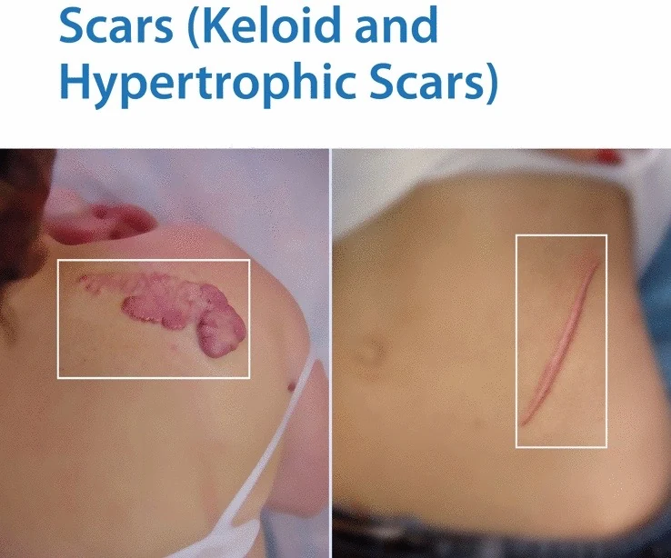
Radiotherapy for Keloid Scarring
Radiotherapy should be delivered post-operatively rather than as monotherapy. Surgical technique must be optimized for best results.
In this article
- Post-excision keloid recurrence
- Evidence for Post-Operative Radiotherapy
- Case series example
- Comparative Study
- Ear keloids: Excision + 5FU/steroid injections vs excision + RT
- RCT: Cryotherapy vs excision + adjuvant Tx
- RCT: Earlobe Keloids Excision + RT vs excision + steroid injections
- Risk-Adapted Post-Operative Radiotherapy
- Risk-Adapted Post-Operative Radiotherapy: Standard Radiotherapy Dosing Schedule
- Risk-Adapted Post-Operative Radiotherapy: Initial Risk Adaptation
- Risk-Adapted Post-Operative Radiotherapy: Surgical modification
- Risk-Adapted Post-Operative Radiotherapy: New RT protocol = fewer fractions
- Radiotherapy Toxicity
- Timing of Radiotherapy Initiation
- Pathway for Post-Operative Keloid Radiotherapy
- Primary Keloid Radiotherapy (without Surgery)
- Cases: Post-Operative Keloid Radiotherapy
- Case 1 – high-risk site
- Case 2 – intermediate risk site
- Case 3: Low risk site
- Summary
- References
Radiotherapy for keloid scarring. ALATRO-IORBC webinar, 25th July 2023. Dr Richard Shaffer, Radiation Oncologist, President of IORBC.
Post-Excision Keloid Recurrence
- Excision alone: 80%
- Excision + steroid injections: 30%
- Excision + Radiotherapy: 5-10%
Sources: Lawrence et al. Ann Plast Surg 1991 Aug; 27: 164-78. Sclafani et al. Dermatol Surg 1996 Jun;22(6):569-74. Ogawa et al. Scars, Burns & Healing 2019 Volume 5: 1–11.
Evidence for Post-Operative Radiotherapy
- Many retrospective case series
- A few small comparative and randomised trials
- One very large comparative cohort study
- Mix of keloids and hypertrophic scars
- Mix of anatomical sites
- Small patient numbers
- Variable RT dose-fractionations
- Unclear outcome measures
- Short follow-up (need at least 12 months, ideally 24 months)
Case Series Example
Patients: n = 80. 35/80 = earlobe keloids. All had previous treatments.
Freedom from recurrence:
- Earlobes: 91% at 1 year, 79%* at 5 years
- Ant chest: 86% at 1 year, 86% at 5 years
Treatment:
- Extralesional excision
- Post-operative RT within 24 hours
- 10 Gy in 1 fraction
- 60 – 100 kV X-rays
*Note: Some earlobes re-pierced. (Ragoowansi et al. Plast Reconstr Surg. 2003 May;111(6):1853-9)
Comparative Study
Emad et al. Med Princ Pract 2010;19:402–405
Study: Prospective non-randomised comparison. Excision + PORT vs. Cryotherapy + steroid injection. n = 26 patients (76 keloids). Median follow-up = 19 months.
Radiotherapy:
- RT started within 48 hours post-op
- 12 Gy in 3 fractions, 1 fraction per week
- 120 kV photons
“Surgery plus immediate radiotherapy was more efficacious and safe than cryotherapy plus intralesional steroid injection” (Emad et al.)
19 Month Recurrence Rate:
- Excision + RT: 18.2%
- Cryo + steroid injections: 28.1%*
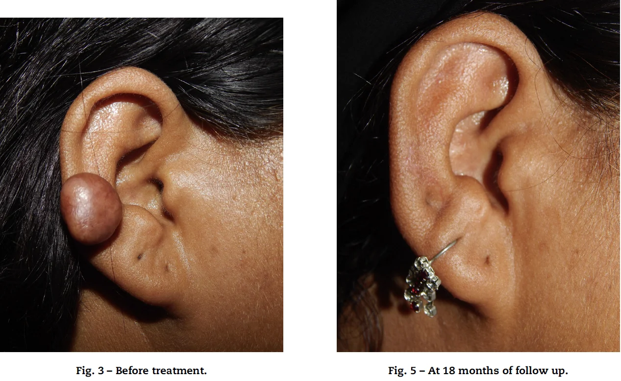
Ear Keloids: Excision + 5FU/steroid Injections vs Excision + RT
Khalid et al. Burns 2018 Sep;44(6):1489-1495
Study: Randomised Controlled Trial. 60 pts, ear keloids. Age = 12 - 65 years. Excision + RT vs 5FU + steroid injections.
Radiotherapy:
- 10 Gy in 2 fractions over 2 days
- Started within 24 hours postop
6 Month Recurrence Rate:
- Excision + RT: 57%
- 5FU + steroid injections: 27%
“Excision with intralesional 5-FU/TAC is more efficacious than excision with radiotherapy in the treatment of ear keloids” (Khalid et al.)
However:
- *Radiotherapy group did much worse than expected
- Electrons to suture line + 5mm margin = much too tight
- Unbalanced groups e.g. more earlobe alone keloids in 5FU group (70% vs 57%)
- Blister/dehiscence in 5FU/TAC group (7%)
- Short follow-up
RCT: Cryotherapy vs Excision + Adjuvant Tx
n = 26. Difficult to recruit. Trial stopped early because poor cryotherapy results.
Two randomisations:
- Primary keloids: Cryotherapy vs excision + steroid injections
- Recurrent keloids: Cryotherapy vs excision + brachytherapy
3 months review: High recurrence in cryotherapy group so stopped recruitment.
“Cryotherapy is inferior to excision + brachytherapy in resistant keloids” (Bijlard et al. Vol 71, Issue 6, p847-856, 2018)
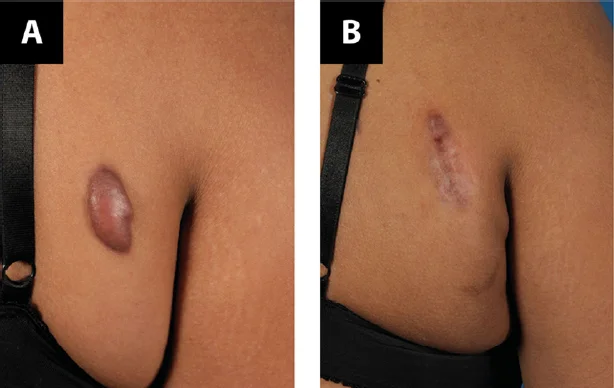
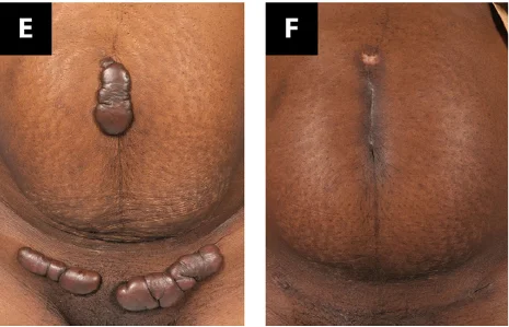
RCT: Earlobe Keloids Excision + RT vs Excision + Steroid Injections
n = 28 keloids. Excised then randomised to: Steroid injections (n=12) vs. Radiotherapy (n=16).
Radiotherapy:
- Randomised to single fraction of 10 Gy (8 keloids) vs 7 Gy (8 keloids)
- 100 kV or electrons
- Within 3 hours postop
Mean follow-up = 18 months.
Recurrence:
- Radiotherapy: 12.5% (2/16)
- Steroid injections: 33% (4/12)
“Further study with larger numbers of patients is warranted to determine if radiation therapy can reduce the risk of recurrence compared with postoperative corticosteroid injections” (Sclafani et al. Dermatol Surg 1996 Jun;22(6):569-74)
Risk-Adapted Post-Operative Radiotherapy
Ogawa et al. (Japan)
Risk-Adapted Post-Operative Radiotherapy: Standard Radiotherapy Dosing Schedule
Up to 2002 (n = 249). Radiotherapy Dose:
- Same to all body sites
- 15Gy in 3 fractions over 3 days (5 Gy per fraction)
- First fraction 24h after surgery
Recurrence:
- Overall = 30%
- Highest = 40% (Chest Wall, scapula, suprapubic, auricle)
- Lowest = 6% (earlobe)
(Ogawa et al. Scars, Burns & Healing 2019 Volume 5: 1–11)
Risk-Adapted Post-Operative Radiotherapy: Initial Risk Adaptation
2003 - 2012 (n = 644). Radiotherapy Dose:
- High-recurrence sites = 20 Gy in 4 fractions
- Intermediate (auricle) = 15 Gy in 3 fractions
- Earlobe = 10 Gy in 2 fractions
Recurrence:
- Overall = 12% (was 30%)
- High rec sites = 13% (was 40%)
- Auricle of ear = 21% (was 40%)
- Earlobe = 7%
(Ogawa et al. Scars, Burns & Healing 2019 Volume 5: 1–11)
Risk-Adapted Post-Operative Radiotherapy: Surgical Modification
2008 - 2012. Surgical modification, RT scheme remained the same:
- Earlobes - wedge excision, simple superficial sutures: RR 6.9% - 4.6%
- Ear auricle – core excision (not wedge): Rec Rate 21.4% → 9%
- High rec sites (Chest Wall, Scapula, Suprapubic): Excision, subcutaneous/fascial tensile reduction sutures and z-plasties. Used for most pts throughout whole 2003-2012 period, so no change in RR
(Ogawa et al. Scars, Burns & Healing 2019 Volume 5: 1–11)
Risk-Adapted Post-Operative Radiotherapy: New RT Protocol = Fewer Fractions
2013 – 2017 (n = 494). Radiotherapy Dose:
- Earlobe: 10Gy/2# → 8Gy in 1 fraction
- High rec sites: 20Gy/4# → 18Gy in 3 fractions
- Other: 15Gy/3# → 15Gy in 2 fractions
Recurrence:
- Overall = 9.3% (in 2002 = 30%)
- High-rec = 10.4% (40%)
- Auricle = 4.5% (21 - 40%)
- Earlobe = 6.7%
(Ogawa et al. Scars, Burns & Healing 2019 Volume 5: 1–11)
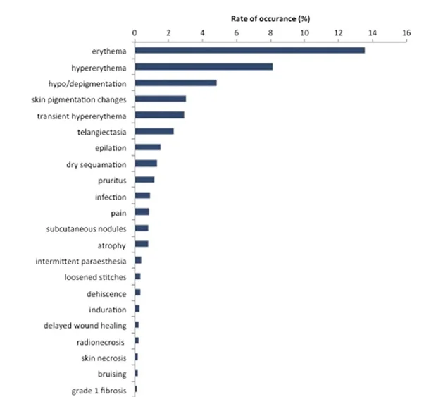
Radiotherapy Toxicity
Acute:
- Skin redness and skin peeling
- Delayed wound healing, dehiscence (< 1%)
Chronic:
- Changes in pigmentation
- Telangectasia – rare, increased with previous steroid injections
- Radiation-induced carcinoma
Radiation-induced carcinoma is dependent on age, body site, OAR volume irradiated, RT dose & technique, e.g. Earlobe keloids = 0.02% lifetime risk of skin cancer (Mankowski et al. 2017).
Timing of Radiotherapy Initiation
- “Many papers state that radiotherapy should be performed immediately after the operation… However, there are also some studies that have found that delaying the start of radiation does not affect the recurrence rate.” (Ogawa et al. Burns & Trauma (2019) 7:39)
- “There was no advantage to beginning radiation therapy within 24 hr following keloidectomy” (Kovalic et al. IJROBP Vol. 17, PP. 77-80) [Mostly earlobe keloids, 75% were within 24 hours postop]
- “A single dose… within the first 72 h after surgical excision of keloid scars appears… highly effective” (Lo et al. Radiot and Onc 1990 Vol 19 p267-272) [Mix of hypertrophic scars and keloids]
Recommendation: Start radiotherapy within 24 - 48 hours after surgery
Pathway for Post-Operative Keloid Radiotherapy
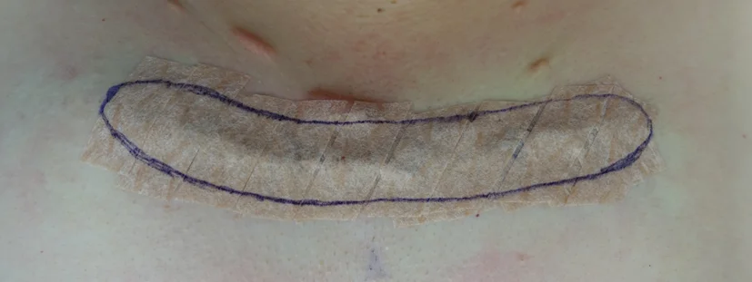
- See Patient In Advance Of Surgery. Surgeon – discuss all options for management, including excision. Radiation Oncologist – history, examination, consent (including RIC)
- Coordinate Dates of Surgery and Radiotherapy
- Surgical excision. Carefully consider dressing presentation
- Radiotherapy markup and delivery
- Superficial-Orthovoltage – simplest, but note depth-dose
- Electrons – consider curvature, junctioning fields
- Brachytherapy – requires infrastructure
- All – consider depth of treatment (Dermo-Epidermal junction)
Primary Keloid Radiotherapy (Without Surgery)
Mankowski (2017):
- Meta-analysis comparing different RT modalities
- Recurrence: 37% after RT monotherapy vs. 22% for post-excision RT
- “Radiation monotherapy of keloids is currently used for very large or nonresectable lesions”
*Malaker (2004):
- Retrospective analysis of 86 keloids treated in 64 patients (1977-2002)
- Dose 37.5 Gy in 5 fractions over 5 weeks (7.5 Gy per fraction)
- 97% showed significant regression at 18 months after the treatment
Darzi (1992):
- 11 keloids and 34 HS treated with RT monotherapy
- 16 Gy in 4 fractions over 2 weeks
- Symptom relief in 27% keloids (65% in hypertrophic scars)
Ogawa:
- 25Gy in 5 fractions (or 24 - 30Gy in 4 - 5 fractions) – anecdotal evidence only
Cases: Post-Operative Keloid Radiotherapy
Case 1 – High-Risk Site
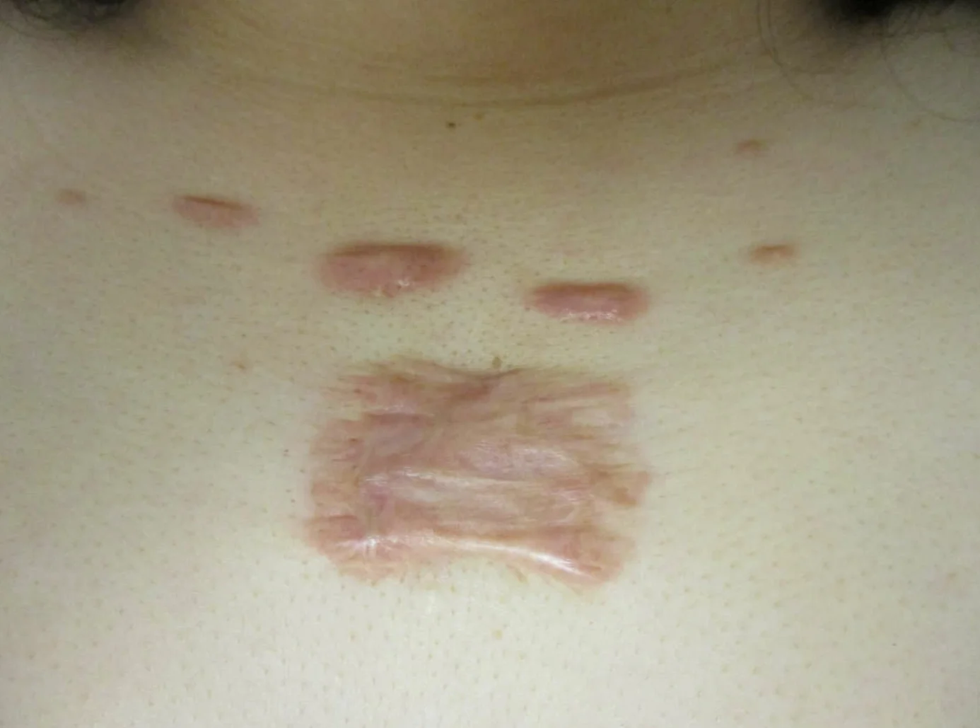
28F, pre-sternal keloid (3.2 x 4.5cm), itchy. Secondary to acne 13y ago. Previous steroid injections, laser.
Treatment:
- Excision + z-plasty
- Post-operative RT (48h post-excision)
- 18Gy in 3 fractions, 6MeV electrons
2 month follow-up.
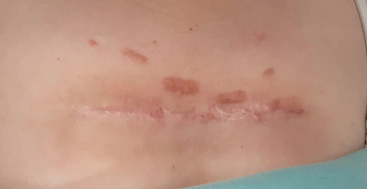
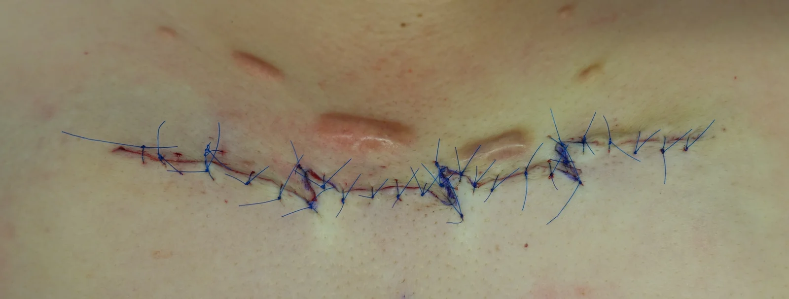
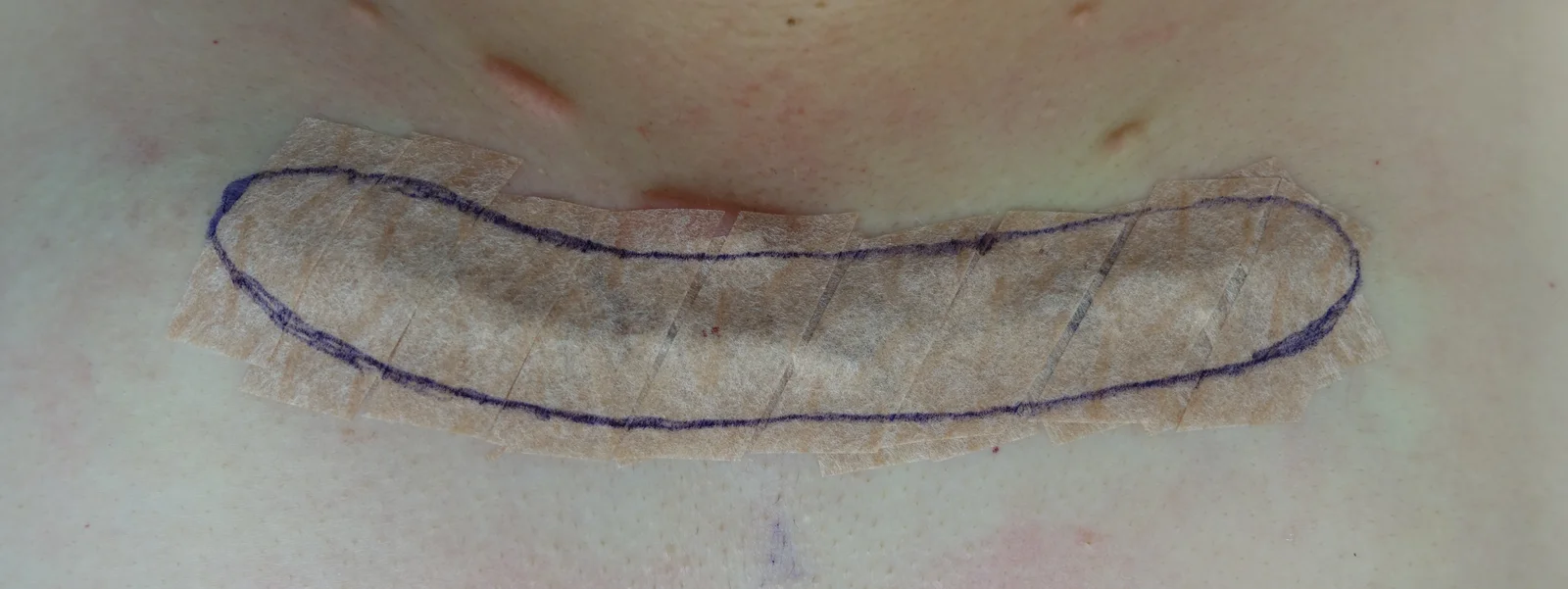
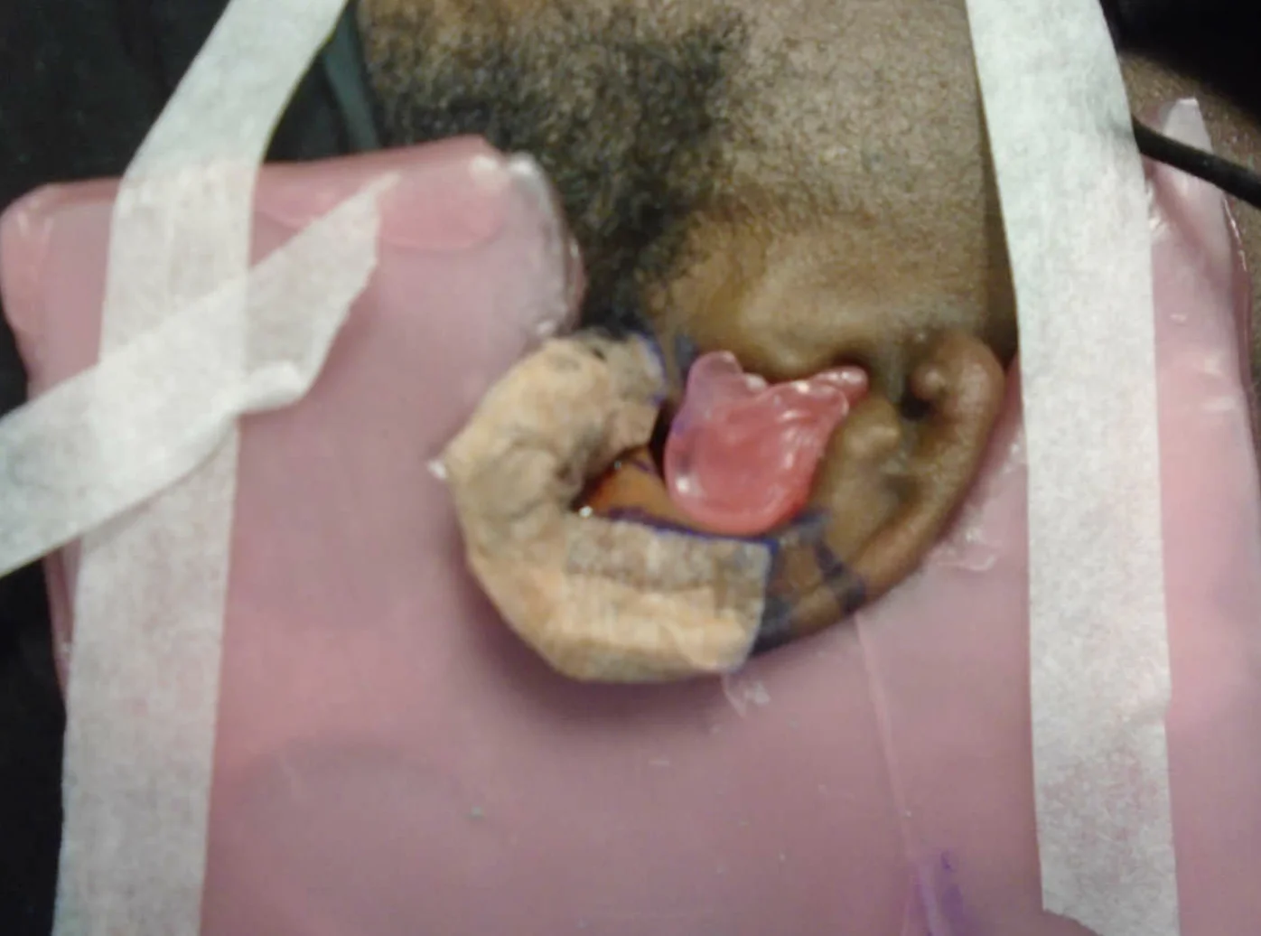
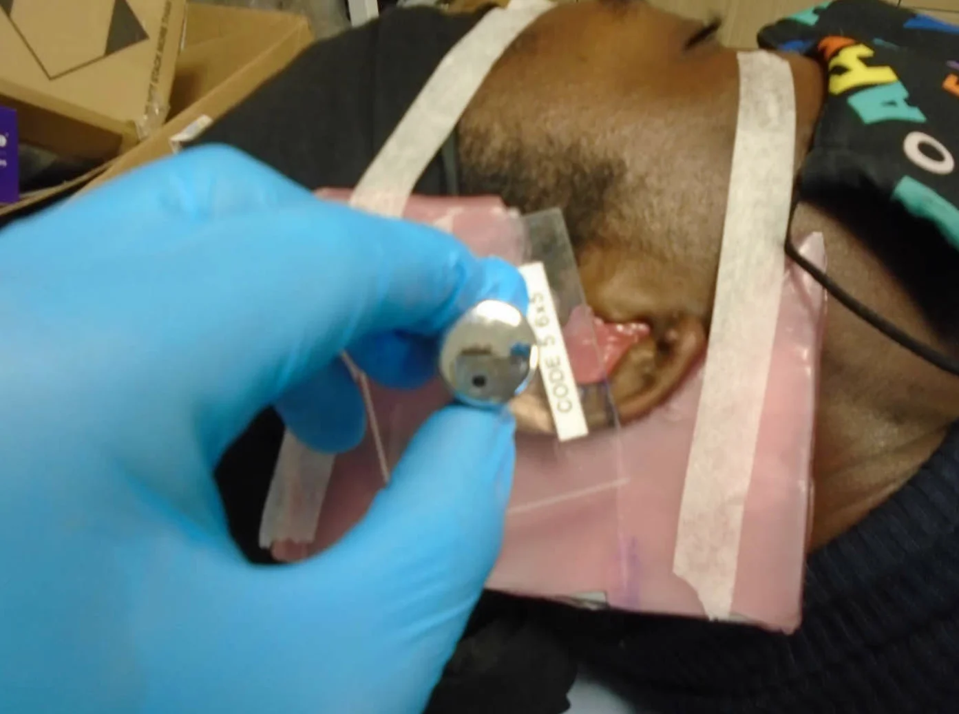
Case 2 – Intermediate Risk Site
23F, bilateral auricle keloids. Due to piercing, increased in size, itchy.
Radiotherapy:
- 24h post-excision
- 15Gy in 2 fractions, electrons
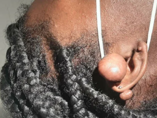
Case 3: Low Risk Site
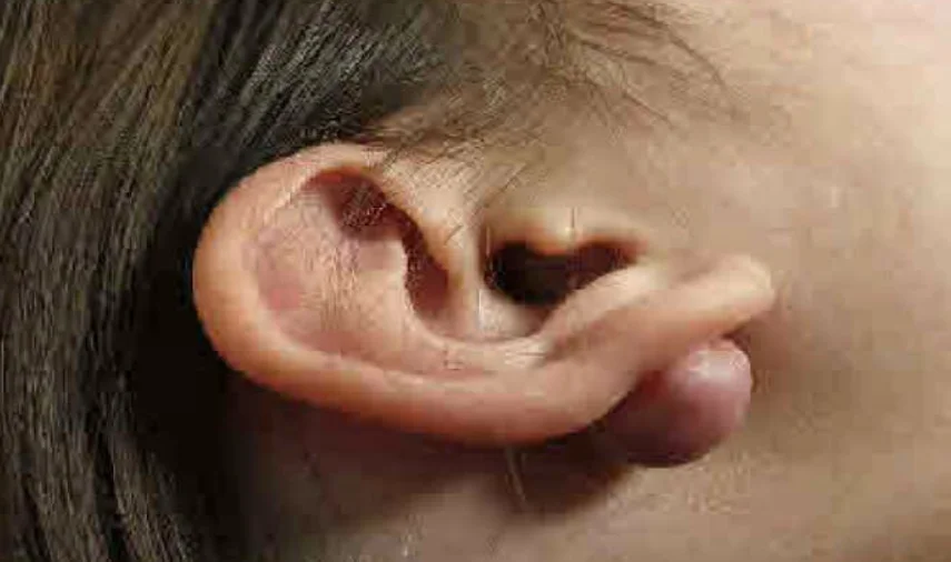
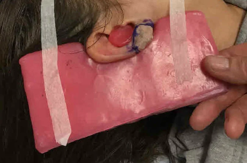
18F, earlobe keloid. Followed piercing. Previous treatment with steroid injections. Radiotherapy = 8Gy in 1 fraction, electrons.
Summary
- Radiotherapy should be delivered post-operatively rather than as monotherapy
- Surgical technique must be optimized for best results
- Start radiotherapy within 24 - 48 hours after excision
- Consider dose adaptation according to anatomical site
- Two year recurrence rate is 5 - 10%
- Toxicity is generally mild
- Radiation-induced carcinoma is very rare but needs careful discussion
References
- Lawrence et al. Ann Plast Surg 1991 Aug; 27: 164-78
- Sclafani et al. Dermatol Surg 1996 Jun;22(6):569-74
- Ogawa et al. Scars, Burns & Healing 2019 Volume 5: 1–11
- Ragoowansi et al. Plast Reconstr Surg. 2003 May;111(6):1853-9
- Emad et al. Med Princ Pract 2010;19:402–405
- Khalid et al. Burns 2018 Sep;44(6):1489-1495
- Bijlard et al. Vol 71, Issue 6, p847-856, 2018
- Ogawa et al. Burns & Trauma (2019) 7:39
- Kovalic et al. IJROBP Vol. 17, PP. 77-80
- Lo et al. Radiot and Onc 1990 Vol 19 p267-272
- Mankowski et al. Optimizing Radiotherapy for Keloids. A Meta-Analysis Systematic Review Comparing Recurrence Rates Between Different Radiation Modalities. Annals of Plastic Surgery Vol 78 (4) 2017
- Malaker et al. Retrospective Analysis of Treatment of Unresectable Keloids with Primary Radiation Over 25 Years. Clinical Oncology (2004) 16: 290–298
- Darzi et al. Evaluation of various methods of treating keloids and hypertrophic scars: a 10-year follow-up study. British Journal of Plastic Surgery (1992), 45, 374-379
Next steps
Thinking About Treatment for Keloids?
Dr Shaffer sees patients for assessment and, where suitable, low-dose radiotherapy.